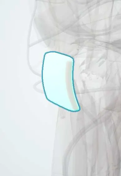
Patellar tendon !
The patellar tendon runs from the kneecap to the shin bone; with the quadriceps it straightens the knee.
Official name: Patellar ligament
Opens in the Durgical app, no sign-up needed.
What it is
A broad, thick band (about 2.5–3 cm wide) from the lower tip of the kneecap to the bump on the front of the tibia (tibial tuberosity).
What it does
Carries the pull of the quadriceps to the shin bone, so the knee can straighten.
Key facts
- Attaches
- Apex and lower margins of patella to tibial tuberosity
- Function
- Continuation of quadriceps tendon; transmits quadriceps pull to tibia to extend knee; tapped for knee jerk (L3–L4)
- Tight in
- Knee flexion; quadriceps contraction
Relations
- Superficial
- Subcutaneous infrapatellar bursa over its lower part and the tibial tuberosity, then skin; the infrapatellar branch of the saphenous nerve crosses it
- Deep
- Infrapatellar (Hoffa) fat pad and, just above its insertion, the deep infrapatellar bursa
- Medial
- Medial retinaculum along its border
- Lateral
- Lateral retinaculum along its border
How to find it
With the knee bent to 90° and relaxed, it is felt as a broad firm band between the lower pole of the patella and the tibial tuberosity; it tightens when the quadriceps contracts.
Examination
- Single-leg decline squat: pain at the lower pole of the patella suggests patellar tendinopathy
- Palpation with the knee straight: tenderness at the lower pole that eases when the knee is flexed is typical of tendinopathy
On imaging
Lateral X-ray: patella alta after rupture; fragments at the tuberosity in Osgood–Schlatter. Ultrasound in tendinopathy: a thickened, darker (hypoechoic) proximal tendon, often with new vessels on Doppler. MRI: thickening and bright signal in the deep proximal fibres.
Common problems
- Tendinitis from jumping and running (jumper's knee).
- Rupture (uncommon): the leg cannot be straightened against gravity.
How it is diagnosed
Physical exam; ultrasound or MRI.
Treatment
- Tendinitis: rest from the painful activity, physical therapy and load management.
- Complete rupture: surgical repair.
- Bone–patellar tendon–bone (BTB) graft: the central third of this tendon, with a small block of bone from the kneecap and the tibia, is a classic graft for ACL reconstruction. It can come from the patient or from a donor.
Clinical notes
- BPTB graft harvest can leave anterior knee pain, pain when kneeling and, rarely, a patellar fracture
- Sinding-Larsen–Johansson disease: traction apophysitis at the lower pole of the patella in adolescents, the upper counterpart of Osgood–Schlatter
- Scarring after surgery or immobilization can shorten the tendon (patella baja), limiting knee motion
Development
The tibial tuberosity ossifies from a tongue-like extension of the proximal tibial epiphysis; repeated pull during the growth spurt causes Osgood–Schlatter disease.
Related conditions
Test yourself
Think of the answer, then tap the question to see it.
Why is the patellar tendon strictly a ligament?
It joins bone to bone (patella to tibial tuberosity), because the patella is a sesamoid bone inside the quadriceps tendon.
Which extensor tendon ruptures typically under 40, and what happens to the patella?
The patellar tendon; the patella rides high (patella alta).
What Insall–Salvati ratios define patella alta and patella baja?
Normal is about 1; above 1.2 is patella alta, below 0.8 patella baja.
Which reflex is tested by tapping the patellar tendon, and at which roots?
The knee jerk, L3–L4.
What is Sinding-Larsen–Johansson disease?
Traction apophysitis at the lower pole of the patella in adolescents, the upper counterpart of Osgood–Schlatter disease.
Educational information, not medical advice. For any symptom, see a health professional.
Its names
- Official Latin (TA2)
- ligamentum patellae TA2 2622
- Official Spanish (DeCS)
- Ligamento rotuliano DeCS D017847
- English (TA2)
- Patellar ligament
- Also called
- infrapatellar portion of quadriceps tendon
Related
Sources: Terminologia Anatomica (TA2); DeCS; Anatomy, Bony Pelvis and Lower Limb: Thigh Quadriceps Muscle.
Spotted a mistake?We are always improving for the community. Tell us what is wrong and our team will review it.Report a mistake
Representative model, for educational purposes only.