FDS !
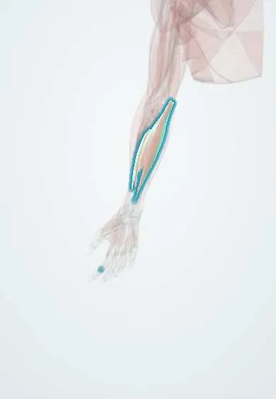
The flexor digitorum superficialis is a forearm muscle with four tendons to the fingers; it bends the fingers at their middle joints.
Official name: Flexor digitorum superficialis
Opens in the Durgical app, no sign-up needed.
What it is
A broad forearm muscle with four tendons to the middle bones of the fingers.
What it does
Bends the fingers at the middle joints.
Key facts
- Origin
- Humero-ulnar head: medial epicondyle and coronoid; radial head: anterior oblique line of the radius
- Insertion
- Sides of the middle phalanges of digits 2–5 (split tendons)
- Action
- Flexes the PIP joints (and MCP joints and wrist)
- Nerve
- Median nerve (C7–T1)
- Blood supply
- Ulnar and radial arteries
Relations
- Superficial
- The superficial flexor layer: pronator teres, flexor carpi radialis, palmaris longus and flexor carpi ulnaris
- Deep
- Flexor digitorum profundus (FDP) and flexor pollicis longus (FPL), with the median nerve held to its deep surface; the ulnar artery passes deep to its upper part
- Course
- Its four tendons pass through the carpal tunnel and enter the fibrous flexor sheaths of the fingers, lying superficial to the FDP tendons until they split
How to find it
Its tendons are felt between palmaris longus and flexor carpi ulnaris in the distal forearm when the fingers bend at the middle joints against resistance; in a forearm exposure it is identified by the median nerve lying under its fascia.
Examination
- FDS test: the other fingers are held straight (blocking the shared FDP belly) and the tested finger is bent; flexion at the PIP joint with a floppy fingertip shows FDS is intact, no flexion means a cut tendon
- In the little finger the test can be falsely positive because its FDS is often weak or tied to the ring finger's
On imaging
Transverse ultrasound shows the FDS tendons in the carpal tunnel superficial to FDP, the middle and ring tendons in front of the index and little. In the finger FDS splits around FDP to reach the middle phalanx. Dynamic scanning during PIP flexion shows gliding or, after a cut, a retracted stump. MRI if unclear; radiographs do not show tendons.
Clinical notes
- Anterior interosseous nerve (AIN) syndrome: the nerve can be squeezed by the FDS arch, the deep head of pronator teres or an accessory FPL head; FPL, FDP to index and middle and pronator quadratus weaken, with no sensory loss
- Flexor zone II ('no man's land', from the distal palmar crease to the FDS insertion): FDS and FDP share a tight sheath, so repairs adhere easily
- The ring finger FDS is a classic donor for opponensplasty and other tendon transfers
Anatomical variants
- The little-finger tendon is weak, absent or linked to the ring finger's in a minority
- Accessory bellies in the palm can present as a mass or crowd the carpal tunnel
Related conditions
Test yourself
Think of the answer, then tap the question to see it.
How is the FDS (flexor digitorum superficialis) tested in a finger?
The other fingers are held straight to block the shared FDP belly; PIP flexion with a floppy fingertip shows the FDS is intact.
What is the Camper chiasm of the FDS?
Each FDS tendon splits over the proximal phalanx to let the FDP tendon through, then inserts on both sides of the middle phalanx.
What is the relationship of the median nerve to the FDS in the forearm?
It passes under the fibrous arch between the FDS heads, then clings to its deep surface down the forearm.
Why do repairs in flexor zone II adhere so easily?
There, from the distal palmar crease to the FDS insertion, the FDS and FDP share a tight sheath.
Educational information, not medical advice. For any symptom, see a health professional.
Its names
- Official Latin (TA2)
- flexor superficialis digitorum TA2 2486
- Official Spanish (DeCS)
- Músculo flexor superficial de los dedos
- English (TA2)
- Flexor digitorum superficialis
- Also called
- flexor digitorum sublimis
Related
- Carpal tunnel
- Median nerve
- Ulnar artery
- Humerus
- Middle phalanx, index finger
- Radius
- Ulna
- Common flexor tendon
Sources: Terminologia Anatomica (TA2); Anatomy, Shoulder and Upper Limb, Hand Flexor Digitorum Superficialis Muscle; Anatomy, Shoulder and Upper Limb, Hand Long Flexor Tendons and Sheaths.
Spotted a mistake?We are always improving for the community. Tell us what is wrong and our team will review it.Report a mistake
Representative model, for educational purposes only.