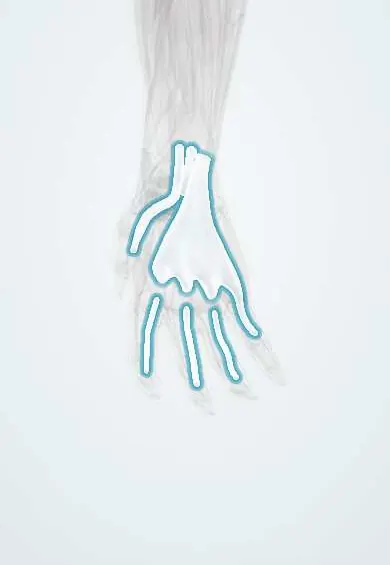
Flexor tendon sheaths !
The flexor tendon sheaths are fluid-lined tubes around the finger flexor tendons; they let the tendons glide, and infection inside them is an emergency.
Official name: Flexor tendon synovial sheaths
Opens in the Durgical app, no sign-up needed.
What it is
Slippery sleeves around the flexor tendons in the wrist, palm and fingers.
What it does
Let the tendons glide; infection inside them spreads fast and is an emergency.
Key facts
- Where
- Carpal tunnel: common flexor sheath (ulnar bursa) and FPL sheath (radial bursa); digital sheaths of fingers 2–4 from the MCP to the DIP
- Function
- Lubricate tendon glide; infection spreads along them (flexor tenosynovitis)
Relations
- Contents
- Ulnar bursa: the FDS and FDP tendons; radial bursa: the FPL tendon; each digital sheath: that finger's FDS and FDP tendons with their vincula
- Superficial
- Flexor retinaculum at the wrist; in the fingers, the fibrous sheath with its annular and cruciate finger pulleys
- Deep
- Carpal bones and palmar wrist ligaments in the carpal tunnel; the volar plates of the finger joints
- Course
- They begin a little above the flexor retinaculum, pass through the carpal tunnel with the median nerve and fan out in the palm
- Above
- Parona space, between the deep flexor tendons and pronator quadratus, into which both bursae can rupture
How to find it
The digital sheaths run along the palmar side of each finger from about the distal palmar crease to the DIP crease; tenderness along this whole line, rather than at one spot, suggests sheath infection.
Examination
- Passive extension of the finger: sharp pain along the sheath is the earliest Kanavel sign and the most useful one in pyogenic flexor tenosynovitis
- Palpation along the whole length of the sheath: tenderness from the fingertip crease to the palm, with a finger held slightly bent and evenly swollen, fits sheath infection; a single tender spot fits a local abscess or a felon
On imaging
Ultrasound shows fluid and thickening within the sheath and helps find a foreign body; MRI maps spread toward the palm and forearm. X-ray looks for a retained foreign body or gas.
Clinical notes
- Pyogenic flexor tenosynovitis is a surgical emergency: late drainage leads to adhesions, tendon necrosis and a stiff finger
- Drainage commonly uses an incision at the A1 pulley and another near the end of the sheath, beyond the DIP, with irrigation of the sheath
- Puncture wounds over the finger creases enter the sheath easily, as the skin there is thin
- Rheumatoid tenosynovitis in the carpal tunnel can compress the median nerve
Anatomical variants
- The little finger sheath usually but not always joins the ulnar bursa; the radial and ulnar bursae are connected in some hands, the route of a horseshoe abscess
Related conditions
Test yourself
Think of the answer, then tap the question to see it.
What are the Kanavel signs, and which is the earliest and most reliable?
Finger held slightly bent, spindle-shaped swelling, tenderness along the sheath and pain on passive extension; pain on passive extension is the earliest and most reliable.
Which digital flexor sheaths reach the wrist, and why does it matter?
The thumb sheath (radial bursa) and the little finger sheath (usually joined to the ulnar bursa), so their infections can spread to the forearm.
What is a horseshoe abscess of the hand?
Infection passing between the thumb and little finger flexor sheaths through a link between the radial and ulnar bursae.
Why is pyogenic flexor tenosynovitis a surgical emergency?
Late drainage leads to adhesions, tendon necrosis and a stiff finger.
Educational information, not medical advice. For any symptom, see a health professional.
Its names
- Official Latin (TA2)
- vagina communis tendinum flexorum TA2 2575
- Official Spanish (DeCS)
- Vainas tendinosas de los flexores (vaina común, vaina del flexor largo del pulgar, vainas sinoviales digitales)
- English (TA2)
- Flexor tendon synovial sheaths
- Also called
- ulnar bursa · radial bursa
Related
- Finger pulleys
- 1st metacarpal
- 5th metacarpal
- Middle phalanx, index finger
- Middle phalanx, middle finger
- Middle phalanx, ring finger
- Proximal phalanx, thumb
- Proximal phalanx, index finger
Sources: Terminologia Anatomica (TA2); Anatomy, Shoulder and Upper Limb, Hand Long Flexor Tendons and Sheaths; Anatomy, Shoulder and Upper Limb, Hand Ulnar Bursa.
Spotted a mistake?We are always improving for the community. Tell us what is wrong and our team will review it.Report a mistake
Representative model, for educational purposes only.