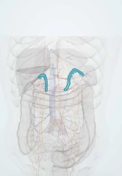
Cardiovascular system · Abdomen
Adrenal arteries !
The suprarenal arteries are many small arteries feeding each adrenal gland, coming from the aorta, the renal artery and the inferior phrenic artery.
Official name: Middle suprarenal artery
Opens in the Durgical app, no sign-up needed.
What it is
Small arteries to the adrenal glands from the inferior phrenic artery, the aorta and the renal artery.
What it does
Supply the adrenal glands.
Key facts
- Origin
- Superior: inferior phrenic artery; middle: aorta (L1); inferior: renal artery
- Branches
- Many small branches (about 50–60) that pierce the capsule of each gland
- Supplies
- Adrenal cortex and medulla; blood leaves by a single suprarenal vein
Relations
- Course
- Superior arteries come down from the inferior phrenic artery, middle arteries pass out from the aorta over the crura, inferior arteries come up from the renal artery; all branch before piercing the capsule
- In front
- Right: inferior vena cava and liver; left: lesser sac, stomach, pancreas and splenic artery
- Behind
- Crura of the diaphragm
- Below
- Upper poles of the kidneys and the renal vessels
How to find it
Not palpable. In adrenalectomy they appear as many small vessels entering the upper, medial and lower edges of the gland from its three sources (inferior phrenic artery, aorta, renal artery); they are sealed as the gland is freed, while the single adrenal vein is found and controlled on its own.
On imaging
Too small for routine CT; adrenal haemorrhage shows as an enlarged, dense gland. Adrenal vein sampling, done through the veins, is used in primary aldosteronism; the short right adrenal vein entering the inferior vena cava makes the right side harder.
Common problems
- Small nodules found by chance on CT (incidentalomas), mostly harmless.
- Tumours that make too much hormone (high blood pressure, Cushing syndrome).
How it is diagnosed
Blood and urine hormone tests; CT or MRI.
Treatment
- Monitoring, medicines, or laparoscopic removal (adrenalectomy).
Clinical notes
- In laparoscopic adrenalectomy the right adrenal vein, short and entering the back of the inferior vena cava, is the main bleeding risk
- Haemorrhage into both adrenal glands can cause acute adrenal insufficiency
Anatomical variants
- The number on each side varies widely; any of the three groups can be multiple or missing
Memory aid
Superior from above (inferior phrenic artery), middle from the middle (aorta), inferior from below (renal artery).
Related conditions
Test yourself
Think of the answer, then tap the question to see it.
What are the three sources of the suprarenal arteries?
Superior from the inferior phrenic artery, middle from the aorta, inferior from the renal artery.
In adrenalectomy, why is the vein, not the arteries, the vessel to control?
Many small arteries (about 50–60) enter each gland and are simply sealed, while blood leaves by a single suprarenal vein.
How does the adrenal blood flow help the medulla make adrenaline?
Blood flows from a subcapsular plexus through the cortex to the medulla, so the medulla receives cortisol at high concentration.
Educational information, not medical advice. For any symptom, see a health professional.
Its names
- Official Latin (TA2)
- arteria suprarenalis media; arteria suprarenalis inferior TA2 4268
- Official Spanish (DeCS)
- Arterias suprarrenales
- English (TA2)
- Middle suprarenal artery
- Also called
- arteria suprarenalis
Related
Sources: Terminologia Anatomica (TA2); Anatomy, Abdomen and Pelvis: Adrenal Glands (Suprarenal Glands); Anatomy, Abdomen and Pelvis: Arteries; Anatomy of the Human Body (20th ed.): The Abdominal Aorta (Lea & Febiger (Gray's Anatomy, 1918), 1918) · adapted, Public domain.
Spotted a mistake?We are always improving for the community. Tell us what is wrong and our team will review it.Report a mistake
Representative model, for educational purposes only.