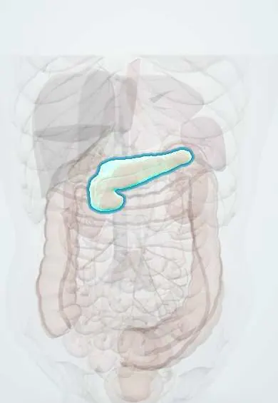
Pancreas !
The pancreas is a long gland behind the stomach that makes digestive juices and the hormones insulin and glucagon, which control blood sugar.
Opens in the Durgical app, no sign-up needed.
What it is
A long gland lying across the back of the upper abdomen, behind the stomach: head (in the curve of the duodenum), body and tail (toward the spleen).
What it does
Makes digestive enzymes and, from tiny islets, the hormones insulin and glucagon that control blood sugar.
Key facts
- Where
- Retroperitoneal across L1–L2: head in duodenal C (uncinate process behind SMV), neck over portal vein, body, tail to splenic hilum
- Blood supply
- Head: superior (gastroduodenal) and inferior (SMA) pancreaticoduodenal arteries; body and tail: splenic artery branches (dorsal, great pancreatic)
- Nerve
- Vagus (secretion) and splanchnic nerves via celiac and superior mesenteric plexuses; pain referred to epigastrium and back
- Function
- Exocrine: digestive enzymes and bicarbonate; endocrine islets: insulin (beta), glucagon (alpha), somatostatin (delta)
Relations
- In front
- Stomach, across the lesser sac; the root of the transverse mesocolon runs along its front
- Behind
- Inferior vena cava and renal veins behind the head; formation of the portal vein behind the neck; abdominal aorta and the origin of the superior mesenteric artery, left kidney and left adrenal gland behind the body
- Lateral
- The head sits in the C-loop of the duodenum, with the common bile duct behind or within it; the tail reaches the splenic hilum in the splenorenal ligament
- Above
- Celiac trunk with the common hepatic artery and the splenic artery, which winds along its upper border
How to find it
Not palpable in health. It is reached by opening the lesser sac through the gastrocolic ligament (part of the greater omentum), by a Kocher maneuver for the head, or by mobilizing the spleen and tail from the left. Its neck projects onto the transpyloric plane (L1).
Examination
- Cullen sign (bruising around the umbilicus) and Grey Turner sign (bruising of the flanks): blood tracking from the retroperitoneum, as in severe hemorrhagic pancreatitis; they appear late
On imaging
Contrast CT (pancreatic protocol) is the key test for complications of pancreatitis and for tumours: necrosis shows as tissue that does not enhance; a low-density mass in the head with a dilated bile duct and pancreatic duct (double duct sign) suggests cancer. Ultrasound looks for gallstones as the cause; endoscopic ultrasound samples small lesions.
Common problems
- Acute pancreatitis, most often from gallstones or alcohol: severe upper abdominal pain spreading to the back.
- Pancreatic cancer.
- Diabetes (islet cells).
How it is diagnosed
Blood tests (lipase), CT, MRI, endoscopic ultrasound.
Treatment
- Hospital care for pancreatitis; remove gallstones; surgery for some tumours.
Clinical notes
- Whipple procedure (pancreaticoduodenectomy): removes the pancreatic head, duodenum, distal common bile duct and gallbladder (often with the distal stomach), then joins the pancreas, bile duct and stomach to the jejunum
- Blunt trauma to the epigastrium (handlebar, steering wheel) can crush the neck against the vertebral column and tear the pancreatic duct
- Pseudocyst: a fluid collection with a fibrous wall, usually in the lesser sac behind the stomach, weeks after pancreatitis
Anatomical variants
- Pancreas divisum: the ducts of the two embryonic buds do not fuse, so most of the gland drains through the minor papilla; the most common congenital variant of the pancreas
- Annular pancreas: a ring of pancreatic tissue around the second part of the duodenum that can block it in newborns (double bubble on X-ray)
- Ectopic pancreatic tissue in the wall of the stomach or duodenum; partial or dorsal agenesis is rare
Development
Forms from dorsal and ventral buds of the foregut endoderm; the ventral bud swings behind the duodenum and fuses with the dorsal bud to make the lower head and uncinate process. Faulty fusion or rotation explains pancreas divisum and annular pancreas.
Memory aid
Causes of acute pancreatitis, 'I GET SMASHED': Idiopathic, Gallstones, Ethanol, Trauma, Steroids, Mumps, Autoimmune, Scorpion sting, Hypercalcemia or Hypertriglyceridemia, ERCP, Drugs.
Related conditions
- Acute pancreatitis
- Chronic pancreatitis
- Pancreatic cancer
- Diabetic ketoacidosis
- Familial chylomicronemia syndrome (lipoprotein lipase deficiency)
- Familial hypertriglyceridaemia
- Hereditary hemochromatosis
- Type 2 diabetes mellitus
- Type 1 diabetes mellitus
- Metabolic syndrome
- Scorpion envenomation (Tityus)
- Hypoglycaemia (including insulin-induced)
- Hyperosmolar hyperglycaemic state
- Insulinoma
- Multiple endocrine neoplasia (MEN 1, 2A, 2B)
- Zollinger-Ellison syndrome (gastrinoma)
- Pancreatic neuroendocrine tumours (VIPoma, glucagonoma, somatostatinoma, non-functioning)
- Gestational diabetes mellitus
- Neonatal hypoglycaemia (including infant of a diabetic mother)
- Cystic fibrosis
Test yourself
Think of the answer, then tap the question to see it.
What are the two main causes of acute pancreatitis?
Gallstones and alcohol; others include very high triglycerides, high calcium, ERCP, trauma and some drugs.
How does pancreatic head cancer classically present?
Painless obstructive jaundice with a palpable gallbladder (Courvoisier sign); body and tail cancers present late with back pain and weight loss.
Which veins form the portal vein behind the neck of the pancreas?
The superior mesenteric vein and the splenic vein.
What does a Whipple procedure remove?
The pancreatic head, duodenum, distal common bile duct and gallbladder, often with the distal stomach.
What is pancreas divisum?
The commonest congenital variant: the ducts of the two buds do not fuse, so most of the gland drains through the minor papilla.
Educational information, not medical advice. For any symptom, see a health professional.
Its names
- Official Latin (TA2)
- pancreas TA2 3114
- Official Spanish (DeCS)
- Páncreas DeCS D010179
- English (TA2)
- Pancreas
Related
Sources: Terminologia Anatomica (TA2); DeCS; Anatomy, Abdomen and Pelvis, Pancreas.
Spotted a mistake?We are always improving for the community. Tell us what is wrong and our team will review it.Report a mistake
Representative model, for educational purposes only.