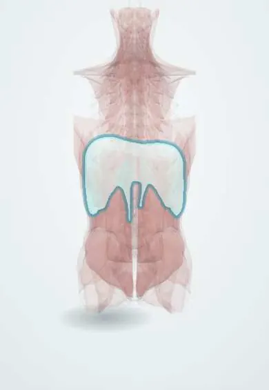
Diaphragm !
The diaphragm is the dome-shaped breathing muscle between the chest and abdomen, anchored to L1 to L3; it is the main muscle for breathing in.
Opens in the Durgical app, no sign-up needed.
What it is
The dome-shaped breathing muscle between the chest and abdomen; its 'crura' attach to the front of L1 to L3.
What it does
The main muscle for breathing in; it also helps raise abdominal pressure to support the spine.
Key facts
- Origin
- Sternal: xiphoid; costal: inner costal cartilages 7–12; lumbar: crura (right L1–L3, left L1–L2) and arcuate ligaments
- Insertion
- Central tendon
- Action
- Main muscle of inspiration (descends, enlarges thorax); raises intra-abdominal pressure
- Nerve
- Phrenic nerve (C3–C5), motor and central sensory; peripheral sensory from lower intercostal nerves
- Blood supply
- Superior and inferior phrenic, pericardiacophrenic, musculophrenic and lower posterior intercostal arteries
Relations
- Above
- Pleura and the bases of both lungs; the pericardium and heart rest on the central tendon
- Below
- Liver on the right; stomach and spleen on the left; the kidneys and suprarenal glands against its posterior part
- Behind
- Crura on the bodies of the upper lumbar vertebrae; the medial and lateral arcuate ligaments arch over psoas major and quadratus lumborum; the sympathetic trunk passes behind the medial arcuate ligament
- Contents
- Caval opening (T8): inferior vena cava; oesophageal hiatus (T10): oesophagus, anterior and posterior vagal trunks, oesophageal branches of the left gastric vessels; aortic hiatus (T12), behind the median arcuate ligament: aorta, thoracic duct, azygos vein
How to find it
It cannot be felt. Its domes are seen on a chest radiograph, the right usually a little higher than the left because of the liver. On ultrasound it is the bright curved line over the liver or spleen that moves down with each breath in; percussing the lower edge of lung resonance in full inspiration and expiration gives a rough measure of its movement.
Examination
- Fluoroscopic sniff test: with a quick sniff a normal dome moves down; a paralysed dome stays still or rises (paradoxical movement), pointing to a phrenic nerve lesion
- Diaphragm ultrasound: a dome that neither descends nor thickens during inspiration supports paralysis on that side
On imaging
Chest radiograph: a raised hemidiaphragm suggests phrenic nerve palsy, lung collapse or a process below it; free gas under the domes on an erect film means a perforated hollow organ. CT shows the crura, the hiatuses and hernia contents.
Clinical notes
- Congenital hernias: Bochdalek (posterolateral, usually left, in newborns, with lung hypoplasia) and Morgagni (anterior, retrosternal, usually right, often found later in life)
- Hiatal hernia through the oesophageal hiatus: the sliding type (the gastro-oesophageal junction moves up) is by far the most common; in the paraoesophageal type the fundus rises beside a junction that stays in place
- Shoulder-tip pain from blood, pus or gas under the diaphragm (ruptured spleen, subphrenic abscess, after laparoscopy) is referred through the phrenic nerve
- Hiccups are involuntary contractions of the diaphragm closed off by the glottis; the reflex runs through the phrenic and vagus nerves, so persistent hiccups can come from irritation anywhere along them
- Phrenic nerve injury (neck or heart surgery, tumour, interscalene block) paralyses one side; paralysis of both sides causes marked breathlessness lying flat
Development
Built from the septum transversum (central tendon), the pleuroperitoneal membranes, the mesentery of the oesophagus (crura) and body-wall muscle. It forms in the neck region and descends, which is why its nerve comes from C3–C5; a pleuroperitoneal membrane that fails to close leaves a Bochdalek hernia.
Memory aid
C3, 4, 5 keeps the diaphragm alive. Openings: I 8 10 Eggs At 12 (IVC T8, oEsophagus T10, Aorta T12).
Related conditions
- Pompe disease (glycogen storage disease type II)
- Respiratory acidosis
- Refeeding syndrome
- Organophosphate and carbamate poisoning
- Coral snake envenomation (Micrurus)
- Oesophageal perforation (Boerhaave syndrome)
- Hiatal hernia
- Trichinellosis
- Critical illness polyneuropathy and myopathy (ICU-acquired weakness)
- Congenital diaphragmatic hernia
- Acute respiratory failure (hypoxaemic and hypercapnic)
- Obesity hypoventilation syndrome
- Penetrating trauma (stab and gunshot wounds)
- Traumatic diaphragmatic injury
- Gastro-oesophageal reflux disease (GERD)
- Achalasia
Test yourself
Think of the answer, then tap the question to see it.
What are the levels of the three main openings of the diaphragm?
Caval T8 (inferior vena cava), oesophageal T10 (oesophagus, vagal trunks), aortic T12 (aorta, thoracic duct, azygos).
What is the motor nerve of the diaphragm, and what are its roots?
The phrenic nerve, C3–C5 («C3, 4, 5 keeps the diaphragm alive»); a complete cord injury above C3 stops spontaneous breathing.
Why does irritation under the central diaphragm cause shoulder-tip pain?
Its central part is sensed by the phrenic nerve (C3–C5), so pain is referred to the C4 dermatome at the shoulder tip.
What does the sniff test show in a paralysed hemidiaphragm?
The paralysed dome stays still or rises (paradoxical movement) instead of descending, pointing to a phrenic nerve lesion.
How do Bochdalek and Morgagni hernias of the diaphragm differ?
Bochdalek: posterolateral, usually left, in newborns with lung hypoplasia. Morgagni: anterior retrosternal, usually right, often found later.
Educational information, not medical advice. For any symptom, see a health professional.
Its names
- Official Latin (TA2)
- diaphragma TA2 2327
- Official Spanish (DeCS)
- Diafragma DeCS D003964
- English (TA2)
- Diaphragm
- Also called
- thoracic diaphragm · respiratory diaphragm · thoracoabdominal diaphragm
Related
Sources: Terminologia Anatomica (TA2); DeCS; Anatomy, Thorax: Diaphragm.
Spotted a mistake?We are always improving for the community. Tell us what is wrong and our team will review it.Report a mistake
Representative model, for educational purposes only.