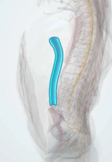
Inferior vena cava !
The inferior vena cava is the body's largest vein, on the front right of the lumbar spine; it returns blood from the legs and abdomen to the heart.
Opens in the Durgical app, no sign-up needed.
What it is
The largest vein in the body, on the front right of the lumbar spine.
What it does
Returns blood from the legs, pelvis and abdomen to the heart. Its wall is thin, so it is handled with great care in anterior spine surgery.
Key facts
- Drains
- Lower limbs, pelvis, kidneys, suprarenals, right gonad, liver (hepatic veins), lumbar veins; formed by the common iliac veins at L5, right of the aorta
- Into
- Right atrium, after passing the caval opening of the diaphragm (T8)
Relations
- Course
- Formed at L5 right of the midline, behind the right common iliac artery; it ascends on the right of the lumbar vertebral bodies, lies in a groove on the back of the liver and passes the caval opening of the diaphragm at T8 to reach the right atrium
- In front
- Right common iliac artery at its start, the root of the mesentery, the third part of the duodenum, the head of the pancreas with the bile duct, the portal vein at the epiploic foramen and the liver
- Behind
- Right side of the lumbar vertebral bodies, the right psoas major, the right sympathetic trunk, the right renal artery and the right crus of the diaphragm
- Medial
- Abdominal aorta, with the right crus between them in the upper abdomen
- Lateral
- Right ureter and the medial border of the right kidney
How to find it
It cannot be felt. In the retroperitoneum it lies on the right side of the lumbar vertebral bodies, to the right of the aorta, from the junction of the common iliac veins at about L5 up to its opening in the diaphragm at T8.
On imaging
Ultrasound below the xiphoid shows it entering the right atrium; it narrows with each breath in (more with a sniff), which helps estimate right atrial pressure. CT shows variants such as a left-sided or duplicated cava.
Clinical notes
- A right-sided exposure of the lumbar spine brings the thin-walled cava into the field; most anterior lumbar approaches are made from the left for this reason
- Obstruction by thrombus, tumour or compression opens collaterals through the ascending lumbar, azygos and hemiazygos veins, the vertebral venous plexuses and the abdominal wall veins
- Kidney cancer can grow along the renal vein into the cava as a tumour thrombus
- Filters can be placed in it, usually below the renal veins, to catch clots from the legs when anticoagulation is not possible
- Behind the liver it lies in a deep groove, sometimes a complete tunnel of liver tissue, and receives the hepatic veins just below the diaphragm; injury of this retrohepatic segment is one of the hardest bleeds to control in liver trauma
Anatomical variants
- Left-sided inferior vena cava, which usually crosses to the right at the level of the left renal vein (rare)
- Duplicated inferior vena cava below the renal veins (rare)
- Azygos continuation: the hepatic segment is absent and the lower body drains through a large azygos vein
Development
It is built from several paired embryonic veins (posterior cardinal, subcardinal and supracardinal) plus a hepatic segment; errors in which parts persist or regress explain its variants.
Memory aid
"Vena cava" has 8 letters, and it passes the diaphragm at T8.
Related conditions
- Hypovolaemic shock
- Neurogenic shock
- Budd-Chiari syndrome
- Dehydration (fluid volume deficit)
- Fluid volume overload (hypervolaemia)
- Renal vein thrombosis
Test yourself
Think of the answer, then tap the question to see it.
Where does the inferior vena cava form, and where does it pass the diaphragm?
It forms at L5 right of the midline from the common iliac veins, and passes the caval opening of the diaphragm at T8.
Why do the left and right gonadal veins drain differently?
The left gonadal vein drains into the left renal vein; the right drains straight into the inferior vena cava.
What is supine hypotensive syndrome of pregnancy, and what relieves it?
The uterus compresses the inferior vena cava when lying flat in late pregnancy; tilting to the left relieves it.
Why is the abdomen left hanging free in prone spine surgery?
Abdominal pressure raises caval pressure and diverts blood into the epidural veins, increasing bleeding.
Educational information, not medical advice. For any symptom, see a health professional.
Its names
- Official Latin (TA2)
- vena cava inferior TA2 4991
- Official Spanish (DeCS)
- Vena cava inferior DeCS D014682
- English (TA2)
- Inferior vena cava
- Also called
- Vena Cava, Inferior
Related
Sources: Terminologia Anatomica (TA2); DeCS; Anatomy, Abdomen and Pelvis: Inferior Vena Cava; Tratado de anatomía humana (9.ª ed.), tomo II, Libro IV Angiología, sección 4: Venas (sistemas de las venas cavas superior e inferior, vena porta, venas del raquis).
Spotted a mistake?We are always improving for the community. Tell us what is wrong and our team will review it.Report a mistake
Representative model, for educational purposes only.