Stomach !
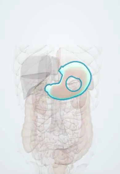
The stomach is the J-shaped bag in the upper left belly, under the ribs, where food is mixed with acid and enzymes before passing to the small intestine.
Opens in the Durgical app, no sign-up needed.
What it is
A J-shaped muscular bag under the left ribs: the cardia, fundus, body and pyloric part (antrum and pylorus).
What it does
Stores food, mixes it with acid and enzymes, and lets it into the duodenum a little at a time.
Key facts
- Where
- Epigastrium and left upper quadrant, intraperitoneal; cardia left of T11, pylorus at L1 (transpyloric plane); cardia, fundus, body, pyloric part
- Blood supply
- Left and right gastric (lesser curvature), right and left gastroepiploic (greater curvature), short gastrics (fundus); all from celiac trunk
- Nerve
- Anterior and posterior vagal trunks (parasympathetic); sympathetic via celiac plexus (T6–T9, greater splanchnic)
- Function
- Food reservoir and mixing; HCl and intrinsic factor (parietal cells), pepsinogen (chief cells), gastrin (antral G cells)
Relations
- In front
- Left lobe of the liver, the diaphragm and the anterior abdominal wall below the left costal margin
- Behind
- The stomach bed, behind the lesser sac: pancreas, splenic artery, left kidney and left suprarenal gland, spleen, transverse mesocolon and the left crus of the diaphragm
- Above
- Left dome of the diaphragm over the fundus; the abdominal esophagus joins it at the cardia, with the vagal trunks on its surface
- Below
- Transverse colon and transverse mesocolon; the greater omentum hangs from the greater curvature
- Lateral
- Spleen, joined to the greater curvature by the gastrosplenic ligament, which carries the short gastric arteries
How to find it
Not palpable in health. Percussion over Traube's space (the lower left anterior chest, over the gastric air bubble) is normally tympanic; dullness there suggests an enlarged spleen or a left pleural effusion. At endoscopy the landmarks are the gastroesophageal junction, the folds of the body, the smooth antrum and the pylorus.
Examination
- Succussion splash: a splashing sound heard over the epigastrium when the abdomen is gently rocked several hours after a meal suggests gastric outlet obstruction
On imaging
An erect chest X-ray shows the gastric air bubble under the left hemidiaphragm, and free air under the diaphragm after perforation (easiest to see on the right, away from the bubble). Endoscopy with biopsy is the main test for ulcers and cancer; CT stages cancer, showing wall thickening, nodes and spread to the omentum and liver.
Common problems
- Gastritis and peptic ulcers, often from H. pylori infection or anti-inflammatory painkillers.
- Reflux (GERD).
- Stomach cancer (more common in parts of Latin America and Asia).
How it is diagnosed
Upper endoscopy with biopsy; H. pylori tests (breath, stool, biopsy).
Treatment
- Acid-reducing medicines, treating H. pylori; surgery for complications or cancer.
Clinical notes
- Signs of advanced gastric cancer: a left supraclavicular node (Virchow node), an umbilical nodule (Sister Mary Joseph nodule) and ovarian spread (Krukenberg tumor)
- Foregut organ: its visceral pain is felt in the epigastrium
- Pyloric stenosis in infants (thickened pyloric muscle) causes projectile non-bilious vomiting and an olive-shaped mass in the epigastrium
- Bariatric operations (sleeve gastrectomy, gastric bypass) reshape it; the vagal trunks at the cardia are at risk in surgery of the gastroesophageal junction
Development
The stomach rotates 90° clockwise around its long axis (seen from above): the left vagus ends up anterior and the right vagus posterior, and the dorsal mesogastrium swings to the left to become the greater omentum, creating the lesser sac behind the stomach.
Memory aid
LARP (English mnemonic): Left vagus Anterior, Right vagus Posterior.
Related conditions
- Peptic ulcer disease
- Upper gastrointestinal bleeding
- Gastric cancer
- Helicobacter pylori gastritis
- Acute (viral) gastroenteritis
- Infantile hypertrophic pyloric stenosis
- Metabolic alkalosis
- Megaloblastic anaemia (B12 or folate deficiency)
- Pernicious anaemia
- Iron deficiency anaemia
- Obesity
- Salicylate (aspirin) toxicity
- Iron poisoning
- Caustic ingestion
- Theophylline toxicity
- Cannabis toxicity and cannabinoid hyperemesis syndrome
- Marine envenomations and seafood poisoning (ciguatera, scombroid, jellyfish, stingray)
- Barrett oesophagus
- Mallory-Weiss syndrome
- Hiatal hernia
- Zollinger-Ellison syndrome (gastrinoma)
- Gastroparesis (delayed gastric emptying)
- Dumping syndrome (rapid gastric emptying)
- Functional dyspepsia
- Ménétrier disease (hypertrophic protein-losing gastropathy)
- Gastrointestinal stromal tumour (GIST)
- Congenital diaphragmatic hernia
- Failure to thrive (faltering growth)
- Traumatic diaphragmatic injury
Test yourself
Think of the answer, then tap the question to see it.
Which arteries supply the stomach?
Left and right gastric (lesser curvature), right and left gastroepiploic (greater curvature) and short gastrics (fundus), all from the celiac trunk.
Which artery can a posterior gastric ulcer erode?
The splenic artery, because the posterior wall faces the lesser sac and stomach bed; a lesser curvature ulcer can erode the left gastric artery.
What sequence links Helicobacter pylori to gastric cancer?
The Correa cascade: gastritis, atrophy, intestinal metaplasia, dysplasia, cancer; gastric cancer is among the leading cancer deaths in Colombia.
Which named signs suggest spread of advanced gastric cancer?
Left supraclavicular node (Virchow node), umbilical nodule (Sister Mary Joseph nodule) and ovarian spread (Krukenberg tumor).
Which cells of the stomach make acid, intrinsic factor and gastrin?
Parietal cells make HCl and intrinsic factor; chief cells pepsinogen; antral G cells gastrin.
Educational information, not medical advice. For any symptom, see a health professional.
Its names
- Official Latin (TA2)
- gaster; fundus gastris; corpus gastris; pars pylorica TA2 2901
- Official Spanish (DeCS)
- Estómago DeCS D013270
- English (TA2)
- Stomach
- Also called
- ventriculus
Related
Sources: Terminologia Anatomica (TA2); DeCS; Anatomy, Abdomen and Pelvis: Stomach.
Spotted a mistake?We are always improving for the community. Tell us what is wrong and our team will review it.Report a mistake
Representative model, for educational purposes only.