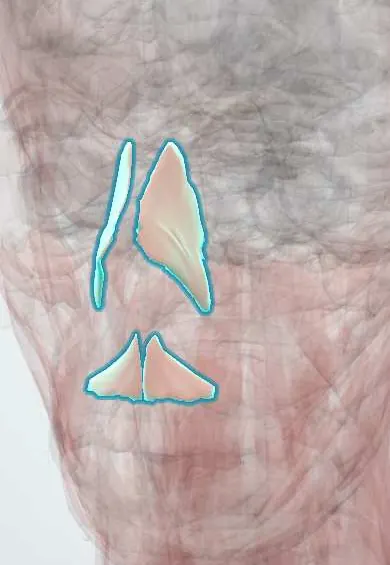
Muscular system · Head and neck
Nasalis !
The nasalis is a group of small muscles over and beside the nose; they narrow and widen the nostrils.
Official name: Nasalis muscle
Opens in the Durgical app, no sign-up needed.
What it is
Small muscles over and under the nose.
What it does
Narrow and widen the nostrils.
Key facts
- Origin
- Maxilla over the canine and lateral incisor
- Insertion
- Nasal cartilages and ala
- Action
- Compresses and dilates the nostrils
- Nerve
- Facial nerve (CN VII): buccal and zygomatic branches
- Blood supply
- Facial artery branches
Relations
- Superficial
- Thin skin and subcutaneous tissue of the nasal dorsum and ala
- Deep
- Upper lateral and alar nasal cartilages, and the maxilla over the canine and lateral incisor at its origin
- Above
- Procerus, joined across the nasal bridge by an aponeurosis
- Lateral
- Levator labii superioris alaeque nasi beside the ala
- Below
- Depressor septi nasi, a small slip from the maxilla to the base of the columella
How to find it
Asked to flare the nostrils, the ala lifts out; asked to wrinkle the nose, short oblique «bunny lines» appear on the side of the nasal bridge over the transverse part.
Examination
- Flaring the nostrils: no movement on one side points to weakness of the facial nerve on that side
On imaging
Not assessed on imaging: on CT of the face it is part of the thin soft tissue over the nasal bones and the alar cartilages. After a blow to the nose, a simple nasal fracture is mainly a clinical diagnosis and radiographs add little; CT is chosen when the injury may extend to the orbit, ethmoid or septum (naso-orbito-ethmoid fracture).
Clinical notes
- Weakness after facial palsy lets the ala collapse on breathing in (external nasal valve collapse)
- Botulinum toxin injection over the transverse part is used for «bunny lines»; spread can reach the lip elevators and change the smile
- Rhinoplasty dissection runs deep to this layer, on the cartilages and bone, to keep the soft tissue envelope intact
Development
Second pharyngeal arch muscle, supplied by the facial nerve like all the muscles of expression.
Test yourself
Think of the answer, then tap the question to see it.
What are the two parts of the nasalis and what does each do?
A transverse part (compressor naris) across the nasal bridge and an alar part (dilator naris) that opens the nostril.
Which nerve supplies the nasalis?
The facial nerve (CN VII), through its buccal (and zygomatic) branches.
What does nostril flaring in a breathless infant reflect, in terms of the nasalis?
The alar part of the nasalis working hard: a sign of increased work of breathing.
Educational information, not medical advice. For any symptom, see a health professional.
Its names
- Official Latin (TA2)
- musculus nasalis TA2 2062
- Official Spanish (DeCS)
- Músculos nasal y depresor del tabique
- English (TA2)
- Nasalis muscle
Related
Sources: Terminologia Anatomica (TA2); Anatomy, Head and Neck: Facial Muscles; Anatomy, Head and Neck, Nose; Anatomy of the Human Body (20th ed.): Development of the Muscles (Lea & Febiger (Gray's Anatomy, 1918), 1918) · adapted, Public domain.
Spotted a mistake?We are always improving for the community. Tell us what is wrong and our team will review it.Report a mistake
Representative model, for educational purposes only.