Skeletal system · Head and neck
Nasal cartilages !
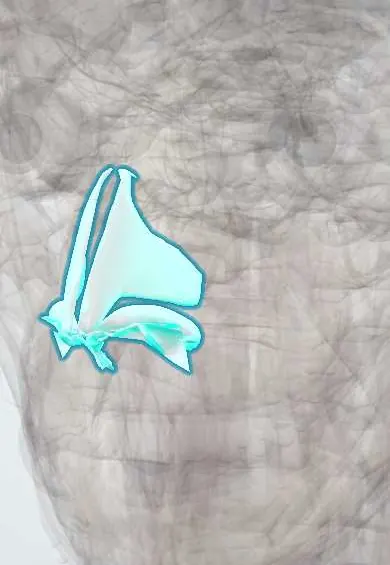
The nasal cartilages are the flexible pieces that shape the tip and sides of the nose and keep the nostrils open.
Official name: Major alar cartilage
Opens in the Durgical app, no sign-up needed.
What it is
The flexible cartilages that shape the tip and sides of the nose.
What it does
Keep the nostrils open.
Key facts
- Attaches
- Lateral nasal and major alar cartilages, joined to the nasal bones, maxillae and septal cartilage
- Function
- Shape the lower nose and keep the nostrils open
Relations
- Above
- Nasal bones, which overlap the upper lateral cartilages at the keystone area
- Medial
- Septal cartilage, fused with the upper lateral cartilages along the dorsum
- Lateral
- Fibrofatty tissue of the ala with small accessory cartilages, and the frontal process of the maxilla at the piriform aperture
- Superficial
- Nasalis and the skin of the nose, thin over the dorsum and thicker and oilier over the tip
- Deep
- Nasal vestibule and the internal and external nasal valves
How to find it
The tip and the alae are felt as springy cartilage below the bony bridge; the domes of the alar cartilages make the tip-defining points, and the medial crura lie in the columella.
Examination
- Cottle test: pulling the cheek outward opens the internal valve; easier breathing suggests valve collapse
- Modified Cottle test: supporting the lateral wall from inside with a probe or cotton bud localises the site of collapse
- Watching the alae during a deep nasal breath shows inspiratory collapse of the external valve
On imaging
Cartilage does not show on radiographs, and nasal bone films add little. CT shows the septum and its deviation, but the alar and upper lateral cartilages blend with the soft tissue of the tip; ultrasound can show them before rhinoplasty. After nasal trauma the key check is clinical: a bulging septal haematoma is drained urgently. CT is kept for suspected facial fractures.
Clinical notes
- Trauma can separate the upper lateral cartilages from the nasal bones or the septum, causing a depression of the middle vault
- Rhinoplasty reshapes the alar cartilages and may add spreader grafts (usually septal cartilage) to widen the internal valve
- A haematoma or infection of the perichondrium can deform the cartilage
Development
In the embryonic face the lateral nasal processes give rise to the upper lateral cartilages and the lateral crura of the alar cartilages, while the medial nasal process forms the columella and the lower septum.
Related conditions
- Basal cell carcinoma
- Rosacea
- Nasal fracture and septal haematoma
- Relapsing polychondritis
- Cleft lip and palate
- Leishmaniasis
Test yourself
Think of the answer, then tap the question to see it.
Where is the internal nasal valve, and why does it matter?
Between the upper lateral cartilage and the septum; it is the narrowest part of the airway, and a narrow angle causes obstruction.
What does the Cottle test assess in the nasal cartilages?
Internal nasal valve collapse: pulling the cheek outward opens the valve, and easier breathing suggests collapse.
Which nasal cartilages shape the tip and nostrils?
The major alar cartilages, with their lateral and medial crura.
Educational information, not medical advice. For any symptom, see a health professional.
Its names
- Official Latin (TA2)
- cartilago alaris major TA2 940
- Official Spanish (DeCS)
- Cartílagos Nasales DeCS D055171
- English (TA2)
- Major alar cartilage
- Also called
- cartilago apicis nasi
Sources: Terminologia Anatomica (TA2); DeCS; Anatomy, Head and Neck, Nose; Anatomy, Head and Neck, Nose Bones; Anatomy of the Human Body (20th ed.): The Branchial Region (Lea & Febiger (Gray's Anatomy, 1918), 1918) · adapted, Public domain.
Spotted a mistake?We are always improving for the community. Tell us what is wrong and our team will review it.Report a mistake
Representative model, for educational purposes only.