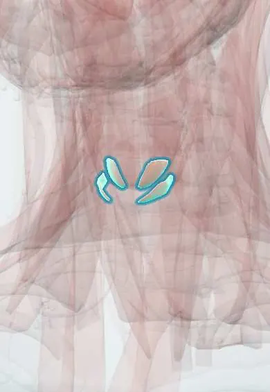
Muscular system · Head and neck
Cricothyroid !
The cricothyroid is a small muscle on the front of the larynx; it tilts the thyroid cartilage to stretch the vocal folds for high notes.
Official name: Oblique part of cricothyroid muscle
Opens in the Durgical app, no sign-up needed.
What it is
A small muscle on the front of the larynx.
What it does
Tilts the thyroid cartilage to stretch the vocal folds for high notes.
Key facts
- Origin
- Anterolateral cricoid arch
- Insertion
- Inferior border and inferior horn of the thyroid cartilage
- Action
- Tilts the thyroid forward: tenses the vocal folds (pitch)
- Nerve
- External branch of the superior laryngeal nerve (X)
- Blood supply
- Superior thyroid artery
Relations
- Superficial
- Sternothyroid muscle and the upper part of the thyroid gland lobe
- Deep
- Cricothyroid joint and the conus elasticus, with the subglottic airway inside
- Medial
- The midline cricothyroid membrane, left bare between the right and left muscles
- Behind
- Inferior pharyngeal constrictor (cricopharyngeal part), which arises just behind it on the side of the cricoid
How to find it
In an anterior neck dissection it is the fan-shaped muscle on the front and side of the cricoid arch, either side of the cricothyroid membrane, under the sternothyroid. On high notes the front gap between the thyroid and cricoid cartilages narrows as it contracts.
Examination
- Laryngoscopy on a rising pitch glide: with one-sided weakness the glottis may look rotated and the affected fold shorter and lower; laryngeal electromyography confirms the nerve injury
On imaging
On axial contrast CT or MRI it is a thin muscle on the front and side of the larynx, between the lower edge of the thyroid cartilage and the cricoid arch. Ultrasound shows the cricothyroid membrane between these cartilages, the landmark for an emergency front-of-neck airway. In superior laryngeal nerve palsy imaging is usually normal; laryngoscopy and laryngeal EMG make the diagnosis.
Clinical notes
- In thyroidectomy the upper-pole vessels are tied close to the gland capsule, where the external laryngeal nerve has usually moved away from the artery
- The external laryngeal nerve can be sought in the sternothyroid–laryngeal (Joll) triangle; intraoperative nerve monitoring helps identify it
- Cricothyroid approximation surgery imitates its pull to raise the pitch of the voice
Anatomical variants
- The crossing point of the external laryngeal nerve with the superior thyroid vessels varies (Cernea types): well above the upper pole, close to it, or below its upper border, the last being most at risk
Development
It comes from the fourth pharyngeal arch, so it is supplied by that arch's nerve, the superior laryngeal branch of the vagus; the other intrinsic laryngeal muscles come from the sixth arch and receive the recurrent laryngeal nerve.
Test yourself
Think of the answer, then tap the question to see it.
What is the action of the cricothyroid muscle?
It tilts the thyroid cartilage forward and down on the cricoid, lengthening and tensing the vocal folds: the main muscle for raising pitch.
What nerve supplies the cricothyroid, and how does it differ from the other intrinsic laryngeal muscles?
The external branch of the superior laryngeal nerve (X); all the other intrinsic laryngeal muscles are supplied by the recurrent laryngeal nerve.
What voice change follows injury to the external laryngeal nerve during thyroidectomy?
A weak, tiring voice without high notes, from cricothyroid paralysis; the nerve runs with the superior thyroid artery.
How are the upper-pole vessels tied in thyroidectomy to protect the nerve to the cricothyroid?
Close to the gland capsule, where the external laryngeal nerve has usually moved away from the superior thyroid artery.
Educational information, not medical advice. For any symptom, see a health professional.
Its names
- Official Latin (TA2)
- pars obliqua musculi cricothyreoidei TA2 2195
- Official Spanish (DeCS)
- Músculo cricotiroideo DeCS D007821
- English (TA2)
- Oblique part of cricothyroid muscle
Related
Sources: Terminologia Anatomica (TA2); DeCS; Anatomy, Head and Neck: Laryngeal Muscles; Anatomy, Head and Neck: Laryngeal Nerves.
Spotted a mistake?We are always improving for the community. Tell us what is wrong and our team will review it.Report a mistake
Representative model, for educational purposes only.