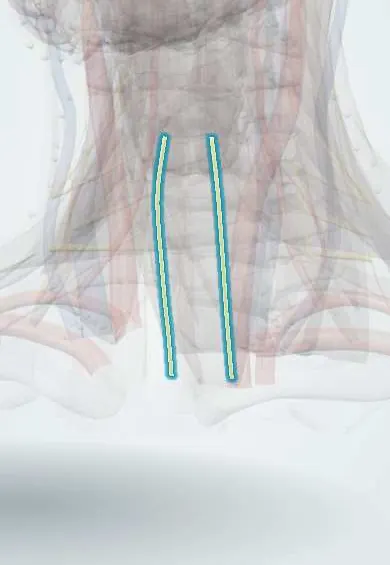
Recurrent laryngeal nerves !
The recurrent laryngeal nerves branch from the vagus in the chest and loop back up to the voice box; damage to them causes hoarseness.
Official name: Recurrent laryngeal nerve
Opens in the Durgical app, no sign-up needed.
What it is
Branches of the vagus that loop back up to the voice box; the left one hooks under the aortic arch.
What it does
Move the vocal cords. Because of its long path, a chest tumour or aortic aneurysm can cause hoarseness.
Key facts
- Roots
- Branch of the vagus; right hooks under the subclavian artery, left under the aortic arch; ascends in the tracheoesophageal groove
- Moves
- All intrinsic laryngeal muscles except cricothyroid; cricopharyngeus
- Feeling from
- Laryngeal mucosa below the vocal folds; trachea and oesophagus
Relations
- Course
- Right: leaves the vagus in front of the right subclavian artery and loops under and behind it. Left: leaves the vagus on the aortic arch and loops under it beside the ligamentum arteriosum. Both climb in the tracheoesophageal groove and enter the larynx below the inferior constrictor
- Medial
- Trachea and esophagus, in the groove between them
- Lateral
- Lobe of the thyroid gland and the inferior thyroid artery, with the parathyroid glands nearby; the common carotid artery further out
- Below
- Left: the aortic arch and ligamentum arteriosum; right: the right subclavian artery
How to find it
In thyroid surgery it is sought in the tracheoesophageal groove where the inferior thyroid artery crosses, and at its entry into the larynx below the inferior constrictor, close to the ligament of Berry; intraoperative nerve monitoring helps identify it.
Examination
- Laryngoscopy (indirect or flexible): the affected vocal fold does not move on phonation and rests near the midline
- Voice and cough assessment: weak, breathy voice, short phonation time and a weak cough on the affected side
On imaging
Not seen itself. After laryngoscopy confirms a vocal fold palsy, CT follows the nerve's course for a cause, on the left down to the aortopulmonary window. CT signs of palsy on that side: the fold sits toward the midline, the laryngeal ventricle widens and the piriform sinus dilates. Neck ultrasound shows thyroid masses beside the tracheoesophageal groove.
Clinical notes
- At risk in thyroid and parathyroid surgery and in anterior approaches to the cervical spine
- The left nerve can be damaged in patent ductus ligation, aortic arch surgery and esophagectomy
- When a vocal fold palsy has no cause in the larynx, imaging covers the whole course of the nerve, down to the aortic arch on the left
Anatomical variants
- Non-recurrent right laryngeal nerve, running straight from the vagus in the neck to the larynx; rare, and almost always with an aberrant right subclavian artery
- The nerve often divides into two or more branches before it enters the larynx
- Its relation to the inferior thyroid artery varies: in front of, behind or between the artery's branches
Development
The loop comes from the 6th pharyngeal arch arteries: on the right the distal part disappears and the nerve moves up to the subclavian artery; on the left it persists as the ductus arteriosus, holding the nerve under the arch.
Related conditions
- Vocal fold paralysis (recurrent laryngeal nerve injury)
- Graves disease (hyperthyroidism)
- Laryngeal cancer
- Thyroid nodule
- Thyroid cancer (papillary)
Test yourself
Think of the answer, then tap the question to see it.
Around which vessels do the right and left recurrent laryngeal nerves hook?
The right under the right subclavian artery; the left under the aortic arch beside the ligamentum arteriosum.
Which laryngeal muscles do the recurrent laryngeal nerves supply?
All intrinsic laryngeal muscles except the cricothyroid.
What is Ortner syndrome?
Hoarseness from compression of the left recurrent laryngeal nerve by an enlarged heart or great vessel, classically the large left atrium of mitral stenosis.
Where is the recurrent laryngeal nerve at risk in thyroid surgery?
In the tracheoesophageal groove where the inferior thyroid artery crosses, and at its entry into the larynx near the ligament of Berry.
How do one-sided and two-sided recurrent laryngeal nerve palsy differ?
One-sided leaves a vocal fold near the midline with a hoarse, breathy voice; two-sided can narrow the airway and cause stridor.
Educational information, not medical advice. For any symptom, see a health professional.
Its names
- Official Latin (TA2)
- nervus laryngeus recurrens TA2 6344
- Official Spanish (DeCS)
- Nervio laríngeo recurrente
- English (TA2)
- Recurrent laryngeal nerve
- Also called
- nervus recurrens
Related
Sources: Terminologia Anatomica (TA2); Anatomy, Head and Neck, Larynx Recurrent Laryngeal Nerve.
Spotted a mistake?We are always improving for the community. Tell us what is wrong and our team will review it.Report a mistake
Representative model, for educational purposes only.