Endocrine system · Head and neck
Thyroid gland !
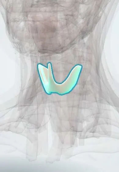
The thyroid gland is a butterfly-shaped gland at the front of the neck below the larynx; it makes hormones that set the body's metabolism.
Opens in the Durgical app, no sign-up needed.
What it is
A butterfly-shaped gland wrapped around the front of the trachea below the larynx.
What it does
Makes thyroid hormones that set the body's metabolism.
Key facts
- Where
- Anterior neck at C5–T1; isthmus over tracheal rings 2–4
- Blood supply
- Superior and inferior thyroid arteries (± thyroid ima)
- Nerve
- Sympathetic (vasomotor); TSH controls secretion
- Function
- T3, T4 (metabolism), calcitonin
Relations
- Superficial
- Infrahyoid (strap) muscles: sternohyoid and sternothyroid, with the sternocleidomastoid further out
- Behind
- Parathyroid glands on the back of each lobe; the recurrent laryngeal nerve in the tracheo-oesophageal groove
- Medial
- Larynx and trachea, bound to it by the suspensory (Berry) ligament; pharynx and oesophagus further back
- Lateral
- Carotid sheath with the common carotid artery, internal jugular vein and vagus nerve
- Above
- Upper poles reach the oblique line of the thyroid cartilage; the external laryngeal nerve runs close to the superior thyroid artery here
- Below
- Lower poles reach about the sixth tracheal ring; a large goitre can extend behind the sternum (retrosternal goitre)
How to find it
From behind the seated person, with the neck slightly bent forward, the fingers rest either side of the trachea below the cricoid cartilage; when the person swallows water, the lobes and any nodule rise under the fingers.
Examination
- Swallowing: a thyroid swelling rises with the larynx; a thyroglossal duct cyst also rises when the tongue is pushed out
- Pemberton sign: raising both arms above the head flushes the face if a retrosternal goitre crowds the thoracic inlet
On imaging
Ultrasound is the first test: size, solid or cystic, margins, calcifications and blood flow of nodules, and guidance for fine-needle aspiration. Radionuclide scans show hot or cold nodules and ectopic thyroid tissue.
Common problems
- Nodules: very common; most are benign.
- Overactive (hyperthyroidism) or underactive (hypothyroidism) gland.
- Goitre: an enlarged gland; thyroid cancer.
How it is diagnosed
Blood tests (TSH, T4), ultrasound and a fine-needle biopsy of suspicious nodules.
Treatment
- Thyroid hormone tablets for hypothyroidism; antithyroid medicines or radioactive iodine for hyperthyroidism.
- Thyroidectomy (removing part or all of the gland) for cancer, large goitres or some hyperthyroidism.
Clinical notes
- Hyperthyroidism (e.g. Graves disease, toxic nodules) and hypothyroidism (e.g. Hashimoto thyroiditis, iodine deficiency) are the two functional extremes
- Thyroidectomy risks: recurrent laryngeal nerve (hoarseness), external laryngeal nerve (loss of high notes), parathyroids (low calcium) and a neck bleed that can press on the airway
- Thyroid veins: the superior and middle veins end in the internal jugular vein; the middle vein is short, has no companion artery and is tied first when the lobe is lifted; the inferior veins form a plexus in front of the trachea that bleeds in a low tracheostomy
Anatomical variants
- Pyramidal lobe rising from the isthmus toward the hyoid bone, a remnant of the thyroglossal duct; common
- Lingual thyroid at the tongue base when descent fails; it may be the only thyroid tissue present
- Thyroid ima artery from the brachiocephalic trunk or aortic arch in a minority, at risk in a low tracheostomy
- Absent isthmus in some people
Development
Endoderm at the foramen caecum descends in front of the hyoid bone and larynx; the calcitonin-making parafollicular (C) cells come from the ultimobranchial body of the fourth pharyngeal pouch.
Related conditions
- Hypothyroidism
- Graves disease (hyperthyroidism)
- Thyroid nodule
- Thyroid cancer (papillary)
- Goitre
- Thyroid storm
- Myxoedema coma
- Hypoparathyroidism (incl. post-thyroidectomy hypocalcaemia)
- Thyroiditis (subacute, painless and postpartum)
- Multiple endocrine neoplasia (MEN 1, 2A, 2B)
- Medullary thyroid carcinoma
- Euthyroid sick syndrome (non-thyroidal illness)
- Follicular, oncocytic (Hürthle cell) and anaplastic thyroid carcinoma
- Autoimmune polyendocrine syndromes (APS-1, APS-2) and IPEX
- Thyroglossal duct cyst and branchial cleft cyst
- Vocal fold paralysis (recurrent laryngeal nerve injury)
- Congenital hypothyroidism
Test yourself
Think of the answer, then tap the question to see it.
Why does a thyroid swelling move up on swallowing?
The thyroid gland is enclosed by the pretracheal fascia and tied to the larynx and trachea, so it rises with the larynx.
How is a thyroglossal duct cyst told apart from other thyroid swellings on examination?
It is a midline lump near the hyoid that also rises when the tongue is pushed out, a remnant of the gland's descent from the foramen caecum.
Which nerve lies behind each thyroid lobe and is the key structure guarded in thyroidectomy?
The recurrent laryngeal nerve, in the tracheo-oesophageal groove near the inferior thyroid artery and Berry ligament; injury causes hoarseness.
What is the Pemberton sign?
Facial flushing when both arms are raised above the head, from a retrosternal goitre crowding the thoracic inlet.
What is the first imaging test for a thyroid nodule?
Ultrasound: size, solid or cystic, margins, calcifications and blood flow, and guidance for fine-needle aspiration.
Educational information, not medical advice. For any symptom, see a health professional.
Its names
- Official Latin (TA2)
- glandula thyreoidea TA2 3863
- Official Spanish (DeCS)
- Glándula Tiroides DeCS D013961
- English (TA2)
- Thyroid gland
Related
- Parathyroid glands
- Recurrent laryngeal nerves
- Trachea
- Cricoid cartilage
- Superior thyroid artery
- Inferior thyroid artery
Sources: Terminologia Anatomica (TA2); DeCS; Anatomy, Head and Neck, Thyroid; Tratado de anatomía humana (9.ª ed.), tomo II, Libro IV Angiología, sección 4: Venas (sistemas de las venas cavas superior e inferior, vena porta, venas del raquis).
Spotted a mistake?We are always improving for the community. Tell us what is wrong and our team will review it.Report a mistake
Representative model, for educational purposes only.