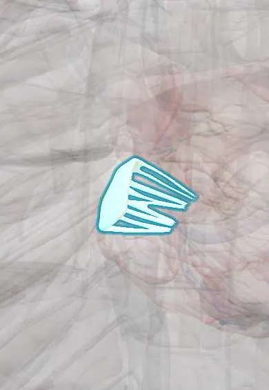
Tricuspid anterior leaflet !
The anterior leaflet is the largest of the three flaps of the tricuspid valve, between the right atrium and ventricle; it closes the front of the opening.
Official name: Superior leaflet of right atrioventricular valve
Opens in the Durgical app, no sign-up needed.
What it is
The largest leaflet of the tricuspid valve, at the front.
What it does
Closes the front part of the valve opening.
Key facts
- Where
- Largest tricuspid leaflet, anterosuperior; valve behind right half of sternum at 4th intercostal space
- Attaches
- Tricuspid annulus; chordae tendineae from anterior and septal papillary muscles
- Function
- Closes in ventricular systole, preventing backflow into right atrium
Relations
- Above
- Supraventricular crest and the infundibulum leading up to the pulmonary valve
- Medial
- Tricuspid septal leaflet, joined at the anteroseptal commissure near the membranous septum
- Below
- Chordae tendineae to the anterior papillary muscle, the largest of the right ventricle papillary muscles; the tricuspid posterior leaflet meets it at the anteroposterior commissure
How to find it
Tricuspid sounds are heard best at the left lower sternal border, 4th to 5th intercostal space. Right-sided murmurs increase with inspiration, which raises venous return to the right heart; severe regurgitation adds large systolic waves in the jugular venous pulse and may make the liver pulsate.
On imaging
Echocardiography: the apical four-chamber view shows the valve with the septal leaflet; the right ventricular inflow and parasternal short-axis views show the anterior leaflet clearly. Colour Doppler grades regurgitation, and the jet velocity estimates right ventricular systolic pressure.
Common problems
- Tricuspid regurgitation, usually because the right ventricle is enlarged (lung or left-heart disease).
- Endocarditis in people who inject drugs.
How it is diagnosed
Echocardiogram.
Treatment
- Treat the cause; surgical repair (ring) during other heart surgery; catheter devices are emerging.
Clinical notes
- Right-sided endocarditis in people who inject drugs most often affects the tricuspid valve; its septic emboli go to the lungs, not the brain
- Ebstein anomaly: the septal and posterior leaflets are displaced toward the apex while the anterior leaflet is large and sail-like
- Pacemaker and defibrillator leads cross the valve and can tether or perforate a leaflet, causing regurgitation
Development
The free leaflets of the atrioventricular valves form where the rim of the atrioventricular canal is undermined and lifted off the ventricular wall. In Ebstein anomaly this separation fails mainly in the septal and posterior leaflets, so the anterior one is spared and large.
Related conditions
Test yourself
Think of the answer, then tap the question to see it.
Which is the largest and most mobile leaflet of the tricuspid valve?
The tricuspid anterior leaflet, which hangs like a curtain between the right atrioventricular opening and the outflow tract.
What murmur does tricuspid regurgitation produce, and what is the Carvallo sign?
A holosystolic murmur at the left lower sternal border; Carvallo sign: it grows louder on inspiration, as venous return to the right heart rises.
Which valve does endocarditis most often affect in people who inject drugs, and where do its emboli go?
The tricuspid valve; its septic emboli go to the lungs, not the brain.
Educational information, not medical advice. For any symptom, see a health professional.
Its names
- Official Latin (TA2)
- cuspis superior valvae atrioventricularis dextrae TA2 3984
- Official Spanish (DeCS)
- Cúspide anterior de la válvula tricúspide DeCS D014261
- English (TA2)
- Superior leaflet of right atrioventricular valve
- Also called
- anterior leaflet of right atrioventricular valve · anterior cusp of right atrioventricular valve · anterosuperior leaflet · cuspis ventralis
Sources: Terminologia Anatomica (TA2); DeCS; Anatomy, Thorax, Heart; Anatomy of the Human Body (20th ed.): Development of the Vascular System (Lea & Febiger (Gray's Anatomy, 1918), 1918) · adapted, Public domain.
Spotted a mistake?We are always improving for the community. Tell us what is wrong and our team will review it.Report a mistake
Representative model, for educational purposes only.