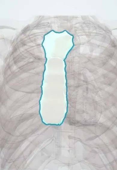
Sternum !
The sternum, or breastbone, is the flat bone in the middle of the chest; it anchors the ribs in front and protects the heart.
Opens in the Durgical app, no sign-up needed.
What it is
The flat breastbone in the middle of the chest: the manubrium on top, the body, and the small xiphoid process at the bottom.
What it does
Anchors the ribs in front and protects the heart. Surgeons split it down the middle to reach the heart (median sternotomy).
Key facts
- Joints with
- Clavicles (sternoclavicular joints); costal cartilages 1–7; manubriosternal and xiphisternal joints
- Landmarks
- Manubrium, body, xiphoid process; jugular notch (T2–T3 level); sternal angle (rib 2, T4–T5 disc level)
Relations
- Behind
- Manubrium: left brachiocephalic vein, the great vessels of the aortic arch and the thymus or its remnant; body: anterior mediastinum, pericardium and the right ventricle
- Lateral
- Costal cartilages 1–7 at the sternocostal joints; the internal thoracic artery and veins run down a little lateral to its borders, behind the cartilages
- Above
- Jugular notch between the sternoclavicular joints, where the clavicles articulate with the manubrium
- Below
- Xiphoid process, with the linea alba and the sternal fibres of the diaphragm attached to it
- In front
- Origin of the pectoralis major on each side; over the midline only skin and thin subcutaneous tissue
How to find it
The jugular notch is felt at the root of the neck. Sliding the finger down a few centimetres, a transverse ridge marks the sternal angle; the 2nd costal cartilage lies just lateral to it, and the 2nd intercostal space below. The body is felt down to the xiphisternal joint, where the xiphoid can be pressed in the epigastric angle.
On imaging
A lateral chest radiograph shows a sternal fracture or displacement best; CT shows fractures, retrosternal haematoma and, after sternotomy, the wires and any separation or collection behind the bone.
Common problems
- Fractures from a steering-wheel or seat-belt impact.
- After open-heart surgery the bone takes 6-8 weeks to heal; wound infection is uncommon but serious.
How it is diagnosed
X-rays or CT.
Treatment
- After a sternotomy the halves are closed with steel wires; patients avoid heavy lifting while it heals.
Clinical notes
- Median sternotomy splits the bone in the midline for heart surgery and is closed with wires; sternal wound infection can extend to the mediastinum (mediastinitis), with diabetes, obesity and harvest of both internal thoracic arteries among the classic risk factors
- Bone marrow can be sampled from the manubrium or upper body in adults; a congenital sternal foramen in the lower body is a hazard for needles, as the heart lies right behind
- Sternal fractures typically follow steering-wheel or seat-belt impact and can occur during CPR; an ECG is usually part of the work-up for associated cardiac contusion
- The xiphoid can be mistaken for an abdominal mass when it is prominent or angled forward
Anatomical variants
- Sternal foramen in the lower body and a bifid or perforated xiphoid process
- Sternalis muscle: a strip running vertically over the sternum in some people, which can mimic a mass on mammography
Development
Forms from two sternal bars that fuse in the midline from above down: incomplete fusion leaves a sternal foramen or, rarely, a sternal cleft. It ossifies from several centres; the xiphoid ossifies last and may stay cartilaginous into adulthood.
Related conditions
- Acute leukaemia (AML and ALL)
- Febrile neutropenia
- Chronic myeloid leukaemia
- Aplastic anaemia
- Polycythaemia vera
- Myelodysplastic syndromes
- Essential thrombocythaemia and primary myelofibrosis
- Sideroblastic anaemia
- Neutropenia and drug-induced agranulocytosis
- Pure red cell aplasia (incl. Diamond–Blackfan anaemia)
- Mediastinitis (acute and fibrosing)
- Blunt cardiac injury
- Flail chest
- Costochondritis
Test yourself
Think of the answer, then tap the question to see it.
Which costal cartilage joins the sternum at the sternal angle, and why does that matter at the bedside?
The 2nd costal cartilage: ribs and intercostal spaces are counted down from it, since the 1st rib is hidden behind the clavicle.
What happens at the level of the sternal angle (T4–T5 disc)?
The aortic arch begins and ends, the trachea divides, and the superior and inferior mediastinum meet.
Where on the sternum are chest compressions given in CPR?
On the lower half of the sternum, in the centre of the chest, above the xiphoid process.
What are the classic risk factors for sternal wound infection after median sternotomy, and where can it spread?
Diabetes, obesity and harvest of both internal thoracic arteries; the infection can extend to the mediastinum (mediastinitis).
Why is a congenital sternal foramen a hazard during sternal bone marrow sampling?
It lies in the lower body of the sternum with the heart right behind it, so a needle can pass through it into the heart.
Educational information, not medical advice. For any symptom, see a health professional.
Its names
- Official Latin (TA2)
- sternum TA2 1129
- Official Spanish (DeCS)
- Esternón DeCS D013249
- English (TA2)
- Sternum
- Also called
- os sternale · sternal bone
Related
Sources: Terminologia Anatomica (TA2); DeCS; Anatomy, Thorax, Sternum; Anatomy, Angle of Louis.
Spotted a mistake?We are always improving for the community. Tell us what is wrong and our team will review it.Report a mistake
Representative model, for educational purposes only.