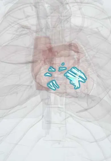
Chordae tendineae !
The chordae tendineae are thin cords inside the ventricles that tie the valve flaps to muscle, stopping them flipping back when the heart squeezes.
Official name: Chordae tendineae of right atrioventricular valve
Opens in the Durgical app, no sign-up needed.
What it is
Thin, strong cords like parachute strings joining the edges of the mitral and tricuspid leaflets to the papillary muscles.
What it does
Stop the leaflets from flipping back into the atria when the ventricles squeeze.
Key facts
- Where
- Fibrous cords in both ventricles
- Attaches
- Papillary muscles (and RV septum) to free edges and ventricular surfaces of tricuspid and mitral leaflets
- Function
- Prevent AV leaflets from everting into atria during systole; rupture causes acute regurgitation
Relations
- Above
- Leaflets: each papillary muscle sends cords to the neighbouring halves of two leaflets, so it holds both sides of one commissure
- Below
- Tips of the left ventricle papillary muscles and right ventricle papillary muscles; on the right some arise straight from the septum
How to find it
In a dissection, opening the ventricle and lifting a leaflet shows the cords fanning out from the papillary tips to the free edge and underside of the leaflet; the two thick strut chords sit on the ventricular face of the anterior mitral leaflet.
On imaging
Echocardiography shows them as fine lines; a ruptured chord appears as a mobile strand whipping into the atrium in systole (flail), best on transesophageal echo. Vegetations may hang from them in endocarditis.
Common problems
- A torn chord makes a leaflet flail and the valve leak suddenly, causing sudden breathlessness.
How it is diagnosed
Echocardiogram.
Treatment
- Surgical repair: resecting the floppy segment or adding artificial chords.
Clinical notes
- Repair replaces torn chordae with artificial cords or transfers healthy ones
- Rheumatic disease thickens, shortens and fuses the chordae, adding narrowing below the valve and making balloon commissurotomy less suitable
Development
They form with the papillary muscles as the ventricular wall is hollowed out: strands of muscle become fibrous cords.
Related conditions
Test yourself
Think of the answer, then tap the question to see it.
How are chordae tendineae classified by where they attach?
First-order reach the free edge and stop prolapse; second-order (including strut chords) reach the ventricular surface; third-order arise from the wall.
What happens when chordae tendineae rupture?
The leaflet becomes flail, with sudden severe regurgitation and lung edema.
Why are the chordae tendineae kept during mitral valve replacement?
Keeping them helps preserve the shape and function of the left ventricle.
How does rheumatic disease change the chordae tendineae?
It thickens, shortens and fuses them, adding narrowing below the valve and making balloon commissurotomy less suitable.
Educational information, not medical advice. For any symptom, see a health professional.
Its names
- Official Latin (TA2)
- chordae tendineae valvae atrioventricularis dextrae; chordae tendineae valvae atrioventricularis sinistrae TA2 4047
- Official Spanish (DeCS)
- Cuerdas tendinosas DeCS D002815
- English (TA2)
- Chordae tendineae of right atrioventricular valve
- Also called
- tendinous cords of right atrioventricular valve · tendinous cords of left atrioventricular valve · tricuspid chordae tendineae
Related
Sources: Terminologia Anatomica (TA2); DeCS; Anatomy, Thorax, Heart Papillary Muscles.
Spotted a mistake?We are always improving for the community. Tell us what is wrong and our team will review it.Report a mistake
Representative model, for educational purposes only.