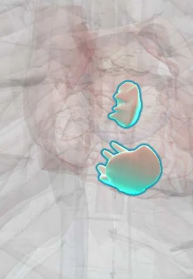
Right ventricle papillary muscles !
The papillary muscles of the right ventricle are small muscle pillars that pull on cords to keep the tricuspid valve closed as the ventricle pumps.
Official name: Papillary muscles of right ventricle
Opens in the Durgical app, no sign-up needed.
What it is
Three small muscles in the right ventricle (anterior, posterior and septal) anchoring the tricuspid chordae.
What it does
Keep the tricuspid valve closed as the right ventricle pumps.
Key facts
- Where
- Right ventricle: anterior (largest, receives moderator band), posterior, and small variable septal muscle
- Attaches
- Ventricular wall to chordae of adjacent tricuspid leaflets
- Function
- Contract in systole, tensing chordae so tricuspid leaflets do not evert
Relations
- In front
- Anterior papillary muscle on the anterior wall, joined by the moderator band from the septum
- Below
- Posterior papillary muscle on the diaphragmatic wall
- Medial
- Septal papillary muscle on the septum, below the anteroseptal commissure
- Above
- Chordae tendineae to the tricuspid anterior leaflet, Tricuspid posterior leaflet and tricuspid septal leaflet
How to find it
Opening the right ventricle through its front wall, the large anterior muscle appears first, joined by the moderator band; the posterior muscle sits on the floor against the diaphragm, and the small septal one high on the septum, below the supraventricular crest.
On imaging
Apical four-chamber echo shows the anterior papillary muscle and the moderator band near the apex; a prominent moderator band is normal and helps identify the morphological right ventricle.
Common problems
- Rarely affected on their own.
How it is diagnosed
Echocardiogram.
Treatment
- Surgical repair: resecting the floppy segment or adding artificial chords.
Clinical notes
- Surgeons use the septal papillary muscle as a landmark for the right bundle branch when repairing ventricular septal defects or the outflow tract
- Leads placed at the right ventricular apex lie among the trabeculae near the anterior papillary muscle and its chordae
- Endomyocardial biopsy, common after heart transplantation, can catch tricuspid chordae or papillary tissue and cause tricuspid regurgitation; the forceps are aimed at the septum to limit this
Anatomical variants
- Their number and size vary; the septal muscle is small, multiple or absent in some hearts
Development
Like the chordae and trabeculae, they are carved out of the spongy muscle meshwork that lines the embryonic ventricle.
Test yourself
Think of the answer, then tap the question to see it.
Which right ventricle papillary muscle receives the moderator band, and why does it contract early?
The anterior papillary muscle, the largest; the moderator band carries the right bundle branch to it as a shortcut.
Why is the septal papillary muscle of the right ventricle a surgical landmark?
It marks where the right bundle branch runs, for repairs of ventricular septal defects or the outflow tract.
What does rupture of a right ventricle papillary muscle cause, and what are its usual causes?
Acute tricuspid regurgitation; it is rare and follows blunt chest trauma or endocarditis.
Educational information, not medical advice. For any symptom, see a health professional.
Its names
- Official Latin (TA2)
- musculi papillares ventriculi dextri TA2 4043
- Official Spanish (DeCS)
- Músculos papilares del ventrículo derecho DeCS D010210
- English (TA2)
- Papillary muscles of right ventricle
Related
Sources: Terminologia Anatomica (TA2); DeCS; Anatomy, Thorax, Heart Papillary Muscles; Anatomy of the Human Body (20th ed.): Development of the Vascular System (Lea & Febiger (Gray's Anatomy, 1918), 1918) · adapted, Public domain.
Spotted a mistake?We are always improving for the community. Tell us what is wrong and our team will review it.Report a mistake
Representative model, for educational purposes only.