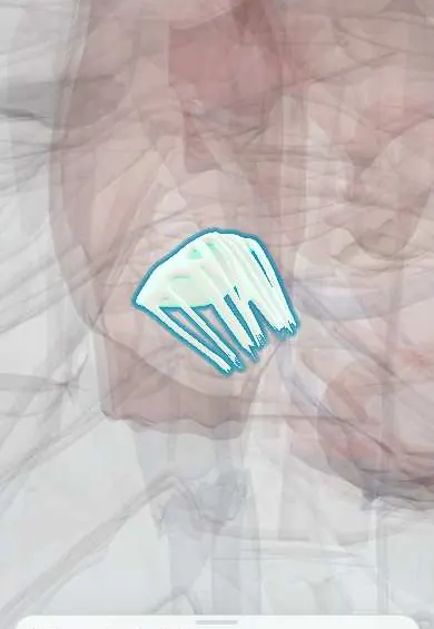
Tricuspid posterior leaflet !
The posterior leaflet is usually the smallest flap of the tricuspid valve, on the lower wall of the right ventricle; it closes the back of the opening.
Official name: Inferior leaflet of right atrioventricular valve
Opens in the Durgical app, no sign-up needed.
What it is
The smallest tricuspid leaflet, at the back (TA2 calls it 'inferior').
What it does
Closes the back part of the valve opening.
Key facts
- Where
- Inferior (posterior) tricuspid leaflet, along diaphragmatic wall of right ventricle; usually smallest
- Attaches
- Tricuspid annulus; chordae tendineae from anterior and posterior papillary muscles
- Function
- Closes in ventricular systole, preventing backflow into right atrium
Relations
- Below
- Diaphragmatic wall of the right ventricle and the Diaphragm beyond; the right coronary artery runs in the atrioventricular groove near its hinge
- In front
- Tricuspid anterior leaflet, joined at the anteroposterior commissure
- Medial
- Tricuspid septal leaflet, joined at the posteroseptal commissure, near the coronary sinus opening in the right atrium
How to find it
In a dissection it is found on the floor of the right ventricle, against the diaphragmatic wall, between the anteroposterior and posteroseptal commissures; its chordae go mainly to the posterior papillary muscle.
On imaging
Echocardiography: best seen in the right ventricular inflow view and the parasternal short axis at valve level; 3D echo shows all three leaflets face-on from the atrium or the ventricle.
Common problems
- Tricuspid regurgitation, usually because the right ventricle is enlarged (lung or left-heart disease).
- Endocarditis in people who inject drugs.
How it is diagnosed
Echocardiogram.
Treatment
- Treat the cause; surgical repair (ring) during other heart surgery; catheter devices are emerging.
Clinical notes
- Ebstein anomaly: displaced septal and posterior leaflet hinges leave an atrialized right ventricle, tricuspid regurgitation and often an accessory pathway (Wolff–Parkinson–White)
- The cavotricuspid isthmus, ablated for typical atrial flutter, reaches the annulus near this leaflet's hinge
- Ring annuloplasty for functional tricuspid regurgitation mainly reduces the free (anterior and posterior) segments of the annulus
Anatomical variants
- Small clefts often split it into two or more scallops, so some texts count four or more tricuspid leaflets
Development
Like the anterior leaflet, it comes from the rim of the atrioventricular canal peeled off the ventricular wall; when it stays stuck down, its hinge appears displaced toward the apex (Ebstein anomaly).
Related conditions
Test yourself
Think of the answer, then tap the question to see it.
Which tricuspid leaflet is usually the smallest, and where does it lie?
The tricuspid posterior (inferior) leaflet, along the diaphragmatic wall of the right ventricle.
In Ebstein anomaly, which tricuspid leaflets are displaced and what results?
The septal and posterior leaflets move toward the apex: an atrialized right ventricle, tricuspid regurgitation and often an accessory pathway (Wolff–Parkinson–White).
Why does the annulus near the tricuspid posterior leaflet stretch in functional tricuspid regurgitation?
Its hinge lies on the free, muscular part of the annulus, which stretches most when the right ventricle dilates.
Educational information, not medical advice. For any symptom, see a health professional.
Its names
- Official Latin (TA2)
- cuspis inferior valvae atrioventricularis dextrae TA2 3985
- Official Spanish (DeCS)
- Cúspide posterior de la válvula tricúspide DeCS D014261
- English (TA2)
- Inferior leaflet of right atrioventricular valve
- Also called
- posterior leaflet of right atrioventricular valve · posterior cusp of right atrioventricular valve · cuspis dorsalis valvulae tricuspidalis
Sources: Terminologia Anatomica (TA2); DeCS; Anatomy, Thorax, Heart; Anatomy of the Human Body (20th ed.): Development of the Vascular System (Lea & Febiger (Gray's Anatomy, 1918), 1918) · adapted, Public domain.
Spotted a mistake?We are always improving for the community. Tell us what is wrong and our team will review it.Report a mistake
Representative model, for educational purposes only.