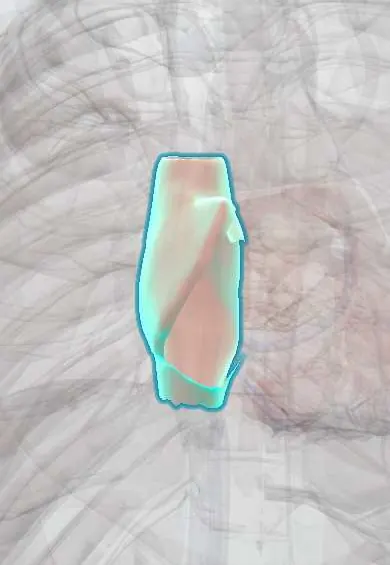
Right atrium !
The right atrium is the upper right chamber of the heart; it collects oxygen-poor blood from the body and passes it to the right ventricle.
Opens in the Durgical app, no sign-up needed.
What it is
The upper right chamber of the heart, which receives blood returning from the body through the venae cavae.
What it does
Collects oxygen-poor blood and pushes it through the tricuspid valve into the right ventricle. The heart's natural pacemaker (the SA node) is in its wall.
Key facts
- Where
- Forms right heart border; receives SVC, IVC and coronary sinus; crista terminalis, pectinate muscles, fossa ovalis
- Blood supply
- Right coronary artery (atrial branches; SA nodal artery in about 60%)
- Nerve
- Cardiac plexus: sympathetic (T1–T4) speeds and strengthens; vagus nerves slow
- Function
- Receives systemic venous blood and fills right ventricle through tricuspid valve; houses SA and AV nodes
Relations
- In front
- Right auricle overlapping the root of the ascending aorta; behind the right sternal border and the right costal cartilages
- Behind
- Right pulmonary veins passing behind it to the left atrium
- Medial
- Interatrial septum with the fossa ovalis, and the left atrium beyond it
- Lateral
- Right lung and mediastinal pleura across the pericardium, with the right phrenic nerve descending on it
- Above
- Superior vena cava entering its upper back part; the Sinoatrial (SA) node lies at this junction, at the top of the crista terminalis
- Below
- Inferior vena cava opening with its small Eustachian valve; the coronary sinus opens just medial to it, in front of the tricuspid valve
How to find it
Its edge projects along the right sternal border, a little outside the sternum, from about the 3rd to the 6th costal cartilage. It has no auscultation area of its own; right atrial pressure is judged at the bedside from the jugular venous pulse in the neck.
On imaging
Frontal chest X-ray: it makes the right heart border, which moves further right when the chamber enlarges. Echocardiography: apical four-chamber and subcostal views; the subcostal view also shows the inferior vena cava and how much it collapses with breathing.
Common problems
- Enlarges when the right side of the heart is under strain (lung disease, tricuspid leak).
- Atrial flutter often circles inside it.
How it is diagnosed
Echocardiogram; ECG for rhythm problems.
Treatment
- Treatment of the underlying cause; catheter ablation for atrial flutter.
Clinical notes
- Central venous catheters and pacing leads reach it from the superior vena cava; catheter tips are usually placed near the cavoatrial junction
- Surgical cannulation or incisions near the superior cavoatrial junction can injure the SA node and cause sinus node dysfunction
- Raised right atrial pressure (right ventricular failure, tricuspid regurgitation, tamponade) shows as distended neck veins and liver congestion
Anatomical variants
- A prominent Eustachian valve or a Chiari network (a lacy remnant of the fetal right venous valve) is a normal finding on echo and must not be mistaken for a clot or vegetation
Development
The smooth part comes from the right horn of the sinus venosus and the rough part from the primitive atrium; the crista terminalis marks where they met. The Eustachian (inferior caval) and Thebesian (coronary sinus) valves are remnants of the right venous valve; in the fetus the Eustachian valve steers caval blood across the foramen ovale.
Related conditions
- Supraventricular tachycardia (AVNRT, AVRT)
- Atrial flutter
- Wolff-Parkinson-White syndrome
- Sinus node dysfunction (sick sinus syndrome) and symptomatic bradycardia
- Restrictive cardiomyopathy (amyloid, sarcoid, haemochromatosis)
- Premature atrial and ventricular contractions
- Tricuspid regurgitation and tricuspid valve disease
- Healthcare-associated infections (CLABSI, CAUTI, VAP)
- Candidiasis: oral, oesophageal and invasive
- Atrial fibrillation
- Cardiac tamponade
- Atrial septal defect
Test yourself
Think of the answer, then tap the question to see it.
Which border of the heart does the right atrium form on a frontal chest X-ray?
The right heart border, between the superior vena cava shadow above and the inferior vena cava below.
What does the crista terminalis separate inside the right atrium?
The smooth posterior sinus venarum from the rough, pectinate-muscle part in front; outside, the sulcus terminalis marks the same line.
What is the ablation target for typical atrial flutter?
The cavotricuspid isthmus of the right atrium, between the inferior vena cava opening and the tricuspid valve.
Why can surgery near the superior cavoatrial junction cause sinus node dysfunction?
The SA node lies at that junction, at the top of the crista terminalis, so cannulation or incisions there can injure it.
Which normal right atrial finding on echo must not be mistaken for a clot or vegetation?
A prominent Eustachian valve or a Chiari network, a lacy remnant of the fetal right venous valve.
Educational information, not medical advice. For any symptom, see a health professional.
Its names
- Official Latin (TA2)
- atrium dextrum; auricula dextra cordis TA2 4022
- Official Spanish (DeCS)
- Atrio derecho DeCS D006325
- English (TA2)
- Right atrium
- Also called
- right atrial appendage
Related
Sources: Terminologia Anatomica (TA2); DeCS; Anatomy, Thorax, Heart; Anatomy, Thorax, Heart and Pericardial Cavity; Tratado de anatomía humana (9.ª ed.), tomo II, Libro IV Angiología, sección 1, capítulo 1: Corazón, artículos I–VI.
Spotted a mistake?We are always improving for the community. Tell us what is wrong and our team will review it.Report a mistake
Representative model, for educational purposes only.