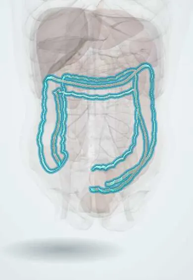
Taeniae coli !
The taeniae coli are three ribbon-like bands of muscle along the colon; they bunch it into pouches and help surgeons tell the colon from the small intestine.
Opens in the Durgical app, no sign-up needed.
What it is
Three ribbons of muscle running along the colon.
What it does
They meet at the base of the appendix, which helps surgeons find it.
Key facts
- Where
- Three bands of longitudinal muscle on cecum and colon (mesocolic, omental, free taenia); absent on appendix and rectum
- Attaches
- Converge at base of appendix; spread into a continuous longitudinal layer at rectosigmoid junction
- Function
- Shorter than the colon wall, so they gather it into haustra; followed to the cecum to locate the appendix
Relations
- In front
- Taenia libera on the ascending, descending and sigmoid colon; on the transverse colon it lies on the inferior surface
- Behind
- Taenia mesocolica, along the attachment of the transverse and sigmoid mesocolon and the fused mesocolon of the ascending and descending colon
- Above
- Taenia omentalis on the transverse colon, where the greater omentum attaches; on the ascending and descending colon it lies posterolaterally
How to find it
Seen at operation as pale, flat ribbons on the colon. At appendectomy the taenia libera is traced down the cecum to the base of the appendix, even when the tip is hidden behind the cecum.
On imaging
Barium enema and CT show the haustral pattern the taeniae create, with folds that only partly cross the lumen; the haustra are lost in long-standing ulcerative colitis.
Common problems
- Colon polyps and colorectal cancer: screening finds them early.
- Diverticular disease and diverticulitis (usually sigmoid).
- Ulcerative colitis and Crohn disease.
How it is diagnosed
Colonoscopy; CT; stool tests for screening.
Treatment
- Polyps removed during colonoscopy; surgery (colectomy) for cancer or complicated disease.
Clinical notes
- The point where the taeniae spread out marks the rectosigmoid junction, which surgeons use to define where the rectum starts
- In sigmoid diverticular disease the taeniae and circular muscle thicken, shortening and narrowing the segment
Related conditions
Test yourself
Think of the answer, then tap the question to see it.
Which features tell the colon from the small intestine at surgery?
Taeniae coli, haustra and fatty epiploic appendices; the appendix and rectum have no taeniae.
How do the taeniae coli help locate the appendix?
They converge on the cecum at the base of the appendix; the taenia libera is traced down even when the tip is hidden.
What does the spreading out of the taeniae coli mark?
The rectosigmoid junction, where they form a continuous longitudinal layer; surgeons use it to define where the rectum starts.
Educational information, not medical advice. For any symptom, see a health professional.
Its names
- Official Latin (TA2)
- taeniae coli TA2 2993
- Official Spanish (DeCS)
- Tenias del colon
- English (TA2)
- Taeniae coli
Sources: Terminologia Anatomica (TA2); Anatomy, Abdomen and Pelvis: Large Intestine.
Spotted a mistake?We are always improving for the community. Tell us what is wrong and our team will review it.Report a mistake
Representative model, for educational purposes only.