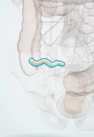
Appendix !
The appendix is a small, finger-like tube attached to the cecum in the lower right belly; when it gets blocked and inflamed, it causes appendicitis.
Official name: Vermiform appendix
Opens in the Durgical app, no sign-up needed.
What it is
A narrow, finger-like tube (about 8-10 cm) hanging from the cecum in the lower right abdomen.
What it does
Its role is unclear; it contains immune tissue and may store helpful gut bacteria.
Key facts
- Where
- Posteromedial cecum about 2 cm below ileocecal junction; retrocecal most common, then pelvic; base under McBurney point
- Blood supply
- Appendicular artery (from ileocolic, SMA) in mesoappendix; an end artery
- Nerve
- Superior mesenteric plexus (T10); visceral pain periumbilical, then parietal pain at McBurney point
- Function
- Gut-associated lymphoid tissue; possible reservoir of gut bacteria
Relations
- In front
- Anterior abdominal wall, greater omentum and loops of ileum
- Behind
- Retrocecal position (the commonest): behind the cecum in the retrocecal recess, on the iliacus and psoas
- Medial
- Pre-ileal and post-ileal positions, in front of or behind the terminal ileum
- Below
- Pelvic position: over the pelvic brim, near the right ovary and uterine tube, the rectum and the bladder
- Contents
- Lymphoid follicles in the mucosa and submucosa, most abundant in childhood and adolescence
How to find it
McBurney point lies one third of the way from the right anterior superior iliac spine to the umbilicus; tenderness there suggests appendicitis. At surgery the taeniae coli are followed down the cecum to its base. In pregnancy the growing uterus pushes the appendix upward and laterally, so pain can be higher.
Examination
- Rovsing sign: pressing the left iliac fossa causes pain in the right iliac fossa
- Psoas sign: pain on passive extension of the right hip (lying on the left side) or on flexing it against resistance; suggests a retrocecal appendix irritating the psoas
- Obturator sign: pain on passive internal rotation of the flexed right hip; suggests a pelvic appendix touching the obturator internus
- Rebound tenderness (Blumberg sign) and guarding mean the parietal peritoneum is inflamed
On imaging
Ultrasound comes first in children and pregnancy: a non-compressible, blind-ending tube wider than about 6 mm, sometimes a fecalith with an acoustic shadow, and bright inflamed fat around it. In adults CT shows a thickened enhancing appendix, fat stranding, fecaliths, abscess or free fluid; MRI is used in pregnancy when ultrasound is inconclusive.
Common problems
- Appendicitis: blocked and infected; pain often starts around the belly button and moves to the lower right, with fever and nausea. If it bursts, peritonitis follows.
How it is diagnosed
Examination, blood tests, ultrasound (children, pregnancy) or CT.
Treatment
- Removal by laparoscopic appendectomy; some uncomplicated cases are treated with antibiotics.
Clinical notes
- Appendectomy: the mesoappendix with the appendicular artery is divided, then the base is tied; the open approach uses a muscle-splitting incision centered on McBurney point
- A retrocecal appendix may give little anterior tenderness and flank pain; a pelvic one may cause diarrhea, urinary frequency and rectal tenderness, delaying the diagnosis
- Perforation is more common in young children and older adults; in small children the short greater omentum walls it off less well
- Neuroendocrine tumors of the appendix are often found by chance in an appendectomy specimen
Anatomical variants
- Tip positions: retrocecal most common, then pelvic; subcecal, pre-ileal and post-ileal less often
- Rare: agenesis or duplication; a left-sided appendix with situs inversus or intestinal malrotation
Development
The cecum and appendix grow from one cecal bud on the midgut loop: its far end lags behind and stays narrow as the appendix while the near end widens into the cecum. In the newborn the appendix sits at the tip of a funnel-shaped cecum; uneven growth of the cecal wall later moves its base to the posteromedial side.
Related conditions
Test yourself
Think of the answer, then tap the question to see it.
Where is McBurney point?
One third of the way from the right anterior superior iliac spine to the umbilicus; it overlies the base of the appendix, and tenderness there suggests appendicitis.
How is the base of the appendix found at surgery?
By following the taeniae coli down the cecum: they converge at the base, which is constant while the tip varies.
What is the most common position of the appendix and which sign does it give?
Retrocecal: little anterior tenderness, flank pain and a psoas sign; a pelvic appendix gives an obturator sign and rectal tenderness.
Why can appendicitis progress quickly to gangrene and perforation?
The appendicular artery (from the ileocolic) is an end artery, so once the blocked lumen swells the blood supply fails.
What usually obstructs the appendix lumen in appendicitis?
A fecalith in adults or lymphoid hyperplasia in children.
Educational information, not medical advice. For any symptom, see a health professional.
Its names
- Official Latin (TA2)
- appendix vermiformis TA2 2976
- Official Spanish (DeCS)
- Apéndice DeCS D001065
- English (TA2)
- Vermiform appendix
- Also called
- processus vermiformis
Related
Sources: Terminologia Anatomica (TA2); DeCS; Anatomy, Abdomen and Pelvis: Appendix; Anatomy of the Human Body (20th ed.): The Digestive Apparatus (Lea & Febiger (Gray's Anatomy, 1918), 1918) · adapted, Public domain.
Spotted a mistake?We are always improving for the community. Tell us what is wrong and our team will review it.Report a mistake
Representative model, for educational purposes only.