Diverticular disease and diverticulitis !
Diverticulitis is inflammation of small pouches (diverticula) in the colon wall, usually in the sigmoid colon; it causes pain in the lower left belly.
Updated 2026-10-29
Structures involved
Tap one to see what it is and open it in 3D.
- Sigmoid colon
- Descending colon
- Taeniae coli
- Mesocolon
- Peritoneum
- Urinary bladder
- Inferior mesenteric artery
- Rectum
In biochemistry
In histology
In the clinical exam
- Abdominal regions and quadrants: what lies under each
- Rebound tenderness (Blumberg sign)
- Acute abdomen and peritoneal irritation
Why it happens
- Patterns of acute inflammation: serous, fibrinous, purulent, abscess, ulcer
- Diet, cancer and systemic disease
- Small bowel and colon motility
- The GI tract wall: mucosa, submucosa, muscularis, serosa; enteric plexuses
What it is
Diverticula are small outpouchings of the colon wall (diverticulosis); diverticulitis is their inflammation or infection, typically in the sigmoid colon, causing left lower quadrant pain.
Who gets it
Diverticulosis becomes more common with age and is frequent in older adults; most people never have symptoms. In Western populations disease is mainly left-sided (sigmoid), whereas right-sided diverticula are more common in Asian populations. Diverticulitis is increasingly seen in younger adults.
Causes
- Herniation of mucosa and submucosa through weak points where the vasa recta penetrate the muscle wall (false diverticula)
- Raised intraluminal pressure and altered colonic motility
- Diverticulitis: microperforation of a diverticulum with local inflammation
Risk factors
- Older age
- Low-fibre, high red meat diet
- Obesity and physical inactivity
- Smoking
- NSAIDs, opioids and corticosteroids (risk of perforation)
- Connective tissue disorders and immunosuppression (complicated disease)
Pathophysiology
High segmental pressure in the sigmoid, the narrowest part of the colon, pushes mucosa out through gaps beside the vessels, between the taeniae coli. Obstruction or erosion of a diverticulum leads to microperforation and pericolic inflammation, which can progress to phlegmon, abscess, free perforation, fistula or stricture.
Classification
- Diverticulosis, symptomatic diverticular disease, diverticular bleeding, diverticulitis
- Uncomplicated vs complicated diverticulitis (abscess, perforation, fistula, obstruction)
- Hinchey classification of perforated disease: I pericolic abscess, II pelvic or distant abscess, III purulent peritonitis, IV faecal peritonitis
- CT-based modified Hinchey or other systems
Symptoms and signs
- Left lower quadrant pain and tenderness, often lasting several days
- Fever and malaise
- Change in bowel habit: constipation or diarrhoea
- Nausea; urinary symptoms if inflammation is next to the bladder
- Palpable tender mass in the left lower abdomen
- Diverticular bleeding: sudden painless rectal bleeding, often without diverticulitis
Diagnosis
- Clinical
- Left lower quadrant pain, tenderness and fever, especially with previous episodes. Examination for peritonitis and a mass; assessment of severity and comorbidities.
- Tests
- Blood count (leucocytosis), CRP, electrolytes, kidney function, lactate if severe; urinalysis; pregnancy test in women of childbearing age.
- Imaging
- CT of the abdomen and pelvis with intravenous contrast confirms the diagnosis, grades severity and shows abscess or free air. Ultrasound is an alternative in some centres. Colonoscopy after recovery (often 6-8 weeks), particularly after complicated disease or if not done recently, to exclude cancer; it is avoided during the acute attack.
Differential diagnosis
- Colorectal cancer (perforated or obstructing)
- Acute appendicitis (including a left-sided appendix or caecal diverticulitis)
- Inflammatory bowel disease, infectious or ischaemic colitis
- Irritable bowel syndrome
- Gynaecological causes: ovarian torsion, ectopic pregnancy, tubo-ovarian abscess
- Kidney stones, pyelonephritis
Goal of treatment
Settle the inflammation and infection, drain or remove complicated disease, avoid unnecessary surgery and antibiotics, and exclude an underlying cancer.
Treatment
- Conservative
- Uncomplicated diverticulitis in selected immunocompetent patients: outpatient care with analgesia and clear fluids then diet as tolerated; antibiotics can be avoided in many cases (guidelines vary).
- Medical
- Antibiotics covering gram-negative and anaerobic bacteria (oral or intravenous) for complicated disease, immunosuppression, sepsis or significant comorbidity; admission when oral intake fails or pain is severe.
- Procedure
- Image-guided percutaneous drainage of larger abscesses, combined with antibiotics; small abscesses often respond to antibiotics alone.
- Surgical
- Emergency surgery for purulent or faecal peritonitis (Hinchey III-IV) or failure of other measures: Hartmann procedure, or resection with primary anastomosis with or without a diverting ileostomy.
- Surgical
- Elective sigmoid resection (often laparoscopic) is individualised: after complicated disease, for fistula or stricture, or with frequent recurrences affecting quality of life.
- Follow-up
- Colonoscopy after recovery to exclude colorectal cancer, and follow-up for recurrence.
Drugs for this condition
Complications
- Abscess (pericolic or pelvic)
- Perforation with purulent or faecal peritonitis
- Fistula, most often colovesical (pneumaturia, faecaluria) or colovaginal
- Stricture and large bowel obstruction
- Recurrence
- Diverticular bleeding (separate from diverticulitis)
Prognosis
Most uncomplicated episodes resolve without surgery, and many patients never have another attack. Complicated disease and perforation with faecal peritonitis carry significant mortality, especially in older and immunosuppressed patients.
Prevention
- High-fibre diet, physical activity and healthy weight
- Not smoking
- Avoiding regular NSAID use when possible
Test yourself
Think of the answer, then tap the question to see it.
Where in the colon does diverticulitis most often occur in Western populations?
The sigmoid colon, causing left lower quadrant pain.
What does Hinchey stage III and IV diverticulitis mean?
Stage III is generalised purulent peritonitis and stage IV is faecal peritonitis; both usually need emergency surgery.
Which imaging test is preferred to diagnose and stage acute diverticulitis?
CT of the abdomen and pelvis with intravenous contrast.
Why is colonoscopy recommended weeks after an episode of diverticulitis, and not during it?
To exclude colorectal cancer, which can mimic diverticulitis; during the acute attack it risks perforation.
Which fistula is the most common complication of diverticulitis, and what are its signs?
A colovesical fistula, which causes pneumaturia, faecaluria and recurrent urinary infections.
Related conditions
- Colorectal cancer
- Acute appendicitis
- Large bowel obstruction
- Peritonitis
- Sigmoid volvulus
- Irritable bowel syndrome
- Kidney stones (urolithiasis)
- Autosomal dominant polycystic kidney disease
Educational information, not medical advice. For any symptom, see a health professional.
Sources: Diverticular Disease of the Colon (StatPearls (NCBI Bookshelf), 2026).
Spotted a mistake?We are always improving for the community. Tell us what is wrong and our team will review it.Report a mistake
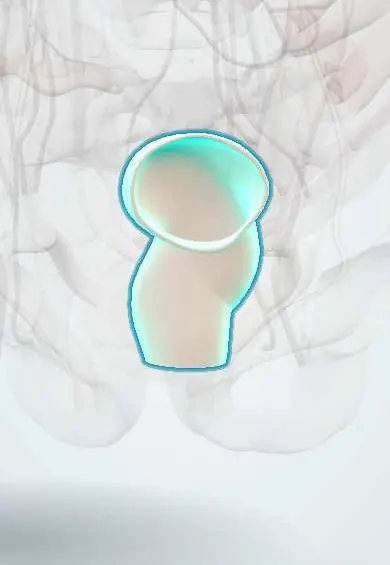
Representative model, for educational purposes only.