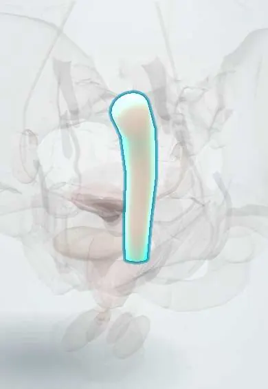
Rectum !
The rectum is the last part of the large bowel, in front of the sacrum and behind the vagina and uterus; it stores stool until the bowel empties.
Opens in the Durgical app, no sign-up needed.
What it is
The last part of the large intestine, behind the vagina and uterus.
What it does
Stores stool until it is time to empty the bowel.
Key facts
- Where
- From S3 to anorectal junction at pelvic floor, about 12–15 cm; peritoneum covers upper third front and sides, middle third front only; no taeniae
- Blood supply
- Superior rectal (IMA), middle rectal (internal iliac) and inferior rectal (internal pudendal) arteries
- Nerve
- Sympathetic: lumbar splanchnics via hypogastric plexuses; parasympathetic: pelvic splanchnic nerves (S2–S4), also carry stretch sensation
- Function
- Stores stool in the ampulla; distension triggers the defecation reflex
Relations
- In front
- Women: rectouterine pouch with loops of small bowel, then the posterior vaginal wall across the rectovaginal septum. Men: rectovesical pouch, then the bladder base, seminal vesicles, ampullae of the vas deferens and prostate
- Behind
- Sacrum and coccyx, the presacral fascia with the presacral venous plexus, median sacral vessels, sacral nodes and sacral nerve roots
- Lateral
- Above, the pararectal fossae of peritoneum; below, the levator ani, the lateral ligaments with the middle rectal vessels, and the inferior hypogastric plexus
- Above
- Rectosigmoid junction, where the taeniae of the sigmoid colon spread into a continuous muscle coat
- Below
- Anorectal junction, pulled forward by the puborectalis, continuing as the anal canal
How to find it
On digital rectal examination the finger passes the anal canal into the lower rectum. Through the front wall it feels the cervix in women, and the prostate (sometimes the seminal vesicles) in men. Above, the rectouterine or rectovesical pouch may be tender (pelvic abscess, peritonitis) or hold a hard shelf (Blumer shelf from peritoneal metastases).
On imaging
Pelvic MRI stages rectal cancer: depth of spread through the wall, nodes, and the distance from the tumour to the mesorectal fascia (circumferential resection margin). Endorectal ultrasound suits early tumours; rigid sigmoidoscopy or MRI measures the height of a tumour from the anal verge.
Common problems
- Rectocele: it bulges into the back wall of the vagina.
- Endometriosis can grow into its wall.
Clinical notes
- In total mesorectal excision the hypogastric nerves and pelvic splanchnic nerves lie just outside the mesorectal fascia; injuring them causes bladder and sexual dysfunction
- Bleeding from the presacral venous plexus during rectal mobilisation can be severe and hard to control
- Haemorrhoids are enlarged vascular cushions of the anal canal; rectal varices are portosystemic collaterals between the superior rectal (portal) and middle and inferior rectal (systemic) veins in portal hypertension: different conditions
- Below the peritoneal reflection the rectum is extraperitoneal, so a full-thickness injury or perforation there does not open into the peritoneal cavity
Anatomical variants
- The number of transverse rectal folds varies, commonly from two to four; the middle fold is the most constant
Development
The urorectal septum divides the cloaca of the hindgut into the rectum and upper anal canal behind and the urogenital sinus in front; faulty division gives anorectal malformations, often with a fistula to the urinary tract or vagina.
Related conditions
- Colorectal cancer
- Ulcerative colitis
- Pelvic organ prolapse
- Obstetric perineal tears and anal sphincter injury
- Haemorrhoids
- Anorectal abscess and fistula
- Rectal prolapse
- Fecal incontinence
- Lower gastrointestinal bleeding
- Colorectal polyps and hereditary polyposis syndromes (FAP, Lynch)
- Constipation and faecal impaction
- Bacterial infectious diarrhoea and dysentery
- Hirschsprung disease
- Anorectal malformations (imperforate anus)
- Functional constipation in children
Test yourself
Think of the answer, then tap the question to see it.
How are the transverse rectal folds (valves of Houston) arranged?
Usually three: upper and lower on the left, middle on the right; the middle fold lies near the anterior peritoneal reflection.
What does total mesorectal excision remove, and why is its plane important?
The rectum within the intact mesorectal fascia; the hypogastric and pelvic splanchnic nerves lie just outside, and injury causes bladder and sexual dysfunction.
Where does lymph from the upper and lower rectum drain?
Upper: along the superior rectal vessels to inferior mesenteric nodes. Lower: also sideways to internal iliac nodes.
How do rectal varices differ from haemorrhoids?
Rectal varices are portosystemic collaterals in portal hypertension; haemorrhoids are enlarged vascular cushions of the anal canal.
What is felt through the front wall of the rectum on digital examination in women and in men?
The cervix in women; the prostate (sometimes the seminal vesicles) in men.
Educational information, not medical advice. For any symptom, see a health professional.
Its names
- Official Latin (TA2)
- rectum TA2 2998
- Official Spanish (DeCS)
- Recto DeCS D012007
- English (TA2)
- Rectum
- Also called
- intestinum rectum · intestinum terminale
Related
Sources: Terminologia Anatomica (TA2); DeCS; Anatomy, Abdomen and Pelvis, Rectum.
Spotted a mistake?We are always improving for the community. Tell us what is wrong and our team will review it.Report a mistake
Representative model, for educational purposes only.