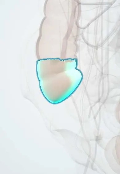
Cecum !
The cecum is the pouch at the start of the large intestine, in the lower right belly, where the small intestine joins and the appendix hangs.
Opens in the Durgical app, no sign-up needed.
What it is
The pouch at the start of the large intestine, in the lower right abdomen, where the ileum enters through the ileocecal valve.
What it does
Receives intestinal contents from the ileum; the appendix hangs from it.
Key facts
- Where
- Right iliac fossa, blind pouch below ileocecal junction; usually intraperitoneal without its own mesentery
- Blood supply
- Anterior and posterior cecal branches of ileocolic artery (SMA)
- Nerve
- Superior mesenteric plexus (sympathetic and vagal); visceral pain referred to periumbilical region (T10)
- Function
- Receives ileal contents through ileocecal valve; absorbs water and salts; appendix opens on its posteromedial wall
Relations
- In front
- Coils of ileum, greater omentum and the anterior abdominal wall of the right iliac fossa
- Behind
- Iliacus and psoas major, with the femoral and lateral femoral cutaneous nerves behind the parietal peritoneum; the retrocecal recess
- Medial
- Terminal ileum entering at the ileocecal valve; the appendix arises below and medial to it
- Above
- Continues as the ascending colon above the level of the ileocecal junction
- Lateral
- Right paracolic gutter
How to find it
It projects onto the right iliac fossa, above the lateral half of the inguinal ligament. At colonoscopy, reaching it is confirmed by seeing the appendiceal orifice, the ileocecal valve and the converging taeniae (the «crow's foot»), sometimes with light shining through the right iliac fossa.
On imaging
On abdominal X-ray and CT a dilated cecum signals distal colonic obstruction or pseudo-obstruction. Cecal volvulus shows a large gas-filled loop often displaced to the upper left abdomen.
Common problems
- Colon polyps and colorectal cancer: screening finds them early.
- Diverticular disease and diverticulitis (usually sigmoid).
- Ulcerative colitis and Crohn disease.
How it is diagnosed
Colonoscopy; CT; stool tests for screening.
Treatment
- Polyps removed during colonoscopy; surgery (colectomy) for cancer or complicated disease.
Clinical notes
- Acute colonic pseudo-obstruction (Ogilvie syndrome) distends mainly the cecum
- Neutropenic enterocolitis (typhlitis): cecal inflammation in patients with very low neutrophil counts, for example during chemotherapy
- Ileocecal tuberculosis and amebic colitis favor this region, both relevant in Latin America
- Pain from the cecum (midgut) is felt around the umbilicus until the overlying parietal peritoneum is involved
Anatomical variants
- A mobile cecum with its own mesentery in some people, which allows cecal volvulus
- A high (subhepatic) cecum from incomplete descent, which moves appendicitis pain upward
Development
The cecum and appendix grow from the cecal bud on the caudal limb of the midgut loop; after the loop returns to the abdomen the cecum descends from below the liver to the right iliac fossa.
Related conditions
- Acute appendicitis
- Colorectal cancer
- Large bowel obstruction
- Crohn disease
- Intussusception (children)
- Lynch syndrome (hereditary non-polyposis colorectal cancer)
- Iron deficiency anaemia
- Soil-transmitted helminthiasis (ascariasis, hookworm, trichuriasis)
- Intestinal amoebiasis (amoebic dysentery)
- Necrotising enterocolitis
Test yourself
Think of the answer, then tap the question to see it.
Why is the cecum the likeliest site of perforation in distal colonic obstruction?
It is the widest part of the colon with the thinnest wall (law of Laplace), especially with a competent ileocecal valve.
How do right-sided colon cancers in the cecum usually present?
With iron-deficiency anemia and weight loss rather than obstruction, because the lumen is wide and the contents liquid.
Which landmarks confirm the cecum is reached at colonoscopy?
The appendiceal orifice, the ileocecal valve and the converging taeniae (the «crow's foot»).
Educational information, not medical advice. For any symptom, see a health professional.
Its names
- Official Latin (TA2)
- caecum; papilla ilealis TA2 2970
- Official Spanish (DeCS)
- Ciego DeCS D002432
- English (TA2)
- Cecum
- Also called
- ileocecal valve · intestinum caecum
Related
Sources: Terminologia Anatomica (TA2); DeCS; Anatomy, Abdomen and Pelvis: Large Intestine.
Spotted a mistake?We are always improving for the community. Tell us what is wrong and our team will review it.Report a mistake
Representative model, for educational purposes only.