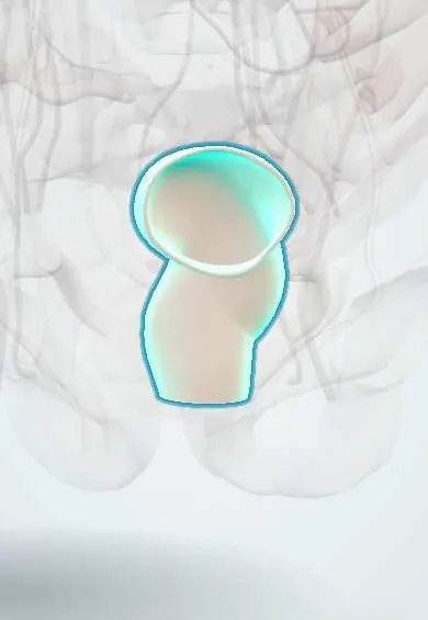
Sigmoid colon !
The sigmoid colon is the S-shaped last part of the colon, in the lower left belly, joining the rectum; it is a common site of diverticula and twisting.
Opens in the Durgical app, no sign-up needed.
What it is
The S-shaped last part of the colon, in the lower left abdomen, joining the rectum.
What it does
Stores stool before it passes to the rectum.
Key facts
- Where
- From pelvic brim to rectosigmoid junction at S3; intraperitoneal on sigmoid mesocolon; S-shaped and mobile
- Blood supply
- Sigmoid arteries (IMA, usually 2–4)
- Nerve
- Inferior mesenteric plexus (sympathetic, L1–L2); pelvic splanchnic nerves (S2–S4)
- Function
- Stores stool before the rectum; common site of diverticula and volvulus
Relations
- Course
- From the pelvic brim it forms an S-shaped loop into the pelvis and back to the midline, joining the rectum where the taeniae spread out and the mesocolon ends
- In front
- Loops of ileum and the anterior abdominal wall of the left iliac fossa
- Behind
- At the apex of its mesocolon: the left ureter, the left common iliac vessels and the gonadal vessels; in the pelvis, the piriformis and the sacral plexus
- Below
- Bladder, and in women the uterus and ovaries; coils sit in the rectovesical or rectouterine pouch
How to find it
It lies in the left iliac fossa and pelvis; in thin people a stool-filled sigmoid is sometimes felt there as a firm cord. Flexible sigmoidoscopy examines it from the anus, for screening and for decompressing a volvulus.
On imaging
Sigmoid volvulus on an abdominal X-ray: the coffee-bean sign, a large inverted-U loop of gas rising from the pelvis toward the upper abdomen; a contrast enema shows a bird-beak taper at the twist. CT is the main test for diverticulitis: fat stranding, wall thickening, abscess or free gas.
Common problems
- Diverticulitis: inflamed pouches; left lower pain and fever.
- Sigmoid volvulus (a twist).
How it is diagnosed
Colonoscopy; CT; stool tests for screening.
Treatment
- Polyps removed during colonoscopy; surgery (colectomy) for cancer or complicated disease.
Clinical notes
- Endoscopic decompression often untwists a volvulus without gangrene; it tends to recur, so a later resection is often planned
- Diverticulitis can form a pericolic abscess or a colovesical fistula (gas or stool in the urine)
- Hartmann procedure: sigmoid resection with an end colostomy and a closed rectal stump, used for perforated diverticulitis or obstruction
- Pain from the sigmoid (hindgut) begins suprapubically and moves to the left iliac fossa when the peritoneum is inflamed, a «left-sided appendicitis» picture
Anatomical variants
- Length and redundancy vary (about 25–40 cm); a long loop with a narrow root predisposes to volvulus
Development
A hindgut derivative that, unlike the descending colon, keeps its mesentery. In Hirschsprung disease neural crest cells fail to reach the end of the bowel, so the rectum and often the sigmoid lack ganglion cells: the newborn passes meconium late and the bowel above the narrow segment distends.
Related conditions
- Colorectal cancer
- Diverticular disease and diverticulitis
- Sigmoid volvulus
- Large bowel obstruction
- Ulcerative colitis
- Irritable bowel syndrome
- Chagas disease
- Lower gastrointestinal bleeding
- Clostridioides difficile infection
- Colorectal polyps and hereditary polyposis syndromes (FAP, Lynch)
- Ischaemic colitis
- Constipation and faecal impaction
- Chagasic megaoesophagus and megacolon
- Bacterial infectious diarrhoea and dysentery
- Hirschsprung disease
- Functional constipation in children
Test yourself
Think of the answer, then tap the question to see it.
What predisposes the sigmoid colon to volvulus?
A long, redundant loop on a narrow mesocolic root; it occurs in older, bedridden or constipated people and in Chagas megacolon.
What does sigmoid volvulus show on a plain abdominal X-ray?
The coffee-bean sign: a large inverted-U loop of gas rising from the pelvis; a contrast enema shows a bird-beak taper.
Why do diverticula form in the sigmoid colon?
The mucosa herniates where vasa recta pierce the circular muscle, between the taeniae; the sigmoid is the commonest site in Western populations.
What is a Hartmann procedure?
Sigmoid resection with an end colostomy and a closed rectal stump, used for perforated diverticulitis or obstruction.
Educational information, not medical advice. For any symptom, see a health professional.
Its names
- Official Latin (TA2)
- colon sigmoideum TA2 2987
- Official Spanish (DeCS)
- Colon sigmoide DeCS D012809
- English (TA2)
- Sigmoid colon
- Also called
- colon sigmoides
Related
Sources: Terminologia Anatomica (TA2); DeCS; Anatomy, Abdomen and Pelvis, Sigmoid Colon; 23.5 The Small and Large Intestines (OpenStax (Rice University), 2022) · adapted, CC BY 4.0.
Spotted a mistake?We are always improving for the community. Tell us what is wrong and our team will review it.Report a mistake
Representative model, for educational purposes only.