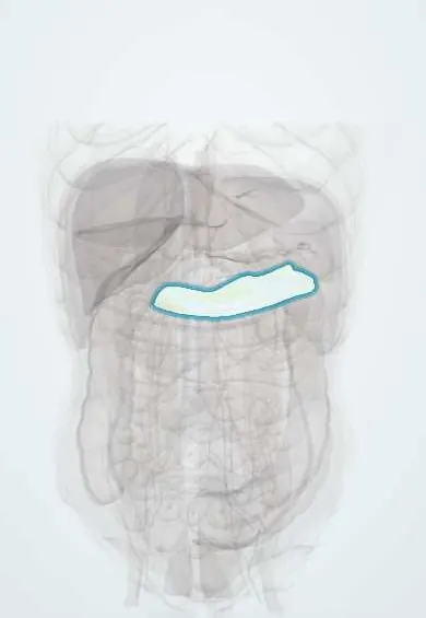
Mesocolon !
The mesocolon is the fold of tissue that attaches parts of the colon (large bowel) to the back wall of the abdomen and carries its vessels and lymph nodes.
Opens in the Durgical app, no sign-up needed.
What it is
Folds of peritoneum attaching parts of the colon (transverse and sigmoid) to the back wall.
What it does
Carry the colic vessels and nodes; in a colectomy the surgeon removes the colon with its mesocolon.
Key facts
- Where
- Transverse mesocolon and sigmoid mesocolon; ascending and descending mesocolon are fused to posterior wall (secondarily retroperitoneal)
- Attaches
- Transverse: root along anterior border of pancreas; sigmoid: inverted-V root with apex over left ureter at common iliac bifurcation
- Function
- Carry colic vessels, nodes and nerves; transverse mesocolon divides supracolic from infracolic compartment
Relations
- Contents
- Colic arteries and veins, the marginal artery, the lymph nodes draining the colon and autonomic nerves
- Behind
- Transverse mesocolon: D2 and the head and body of the pancreas at its root; sigmoid mesocolon: left ureter, iliac vessels and gonadal vessels
- Above
- Above the transverse mesocolon: the lesser sac and the stomach; it forms part of the floor of the lesser sac
- Below
- Below the transverse mesocolon: the small intestine in the infracolic compartment
How to find it
At laparotomy the transverse colon is lifted to show the transverse mesocolon from below with the middle colic vessels; the sigmoid loop is lifted to show its mesocolon and the intersigmoid recess.
On imaging
On CT the mesocolon is fat crossed by the colic vessels; stranding there follows colitis, diverticulitis or pancreatitis, and a whirl of twisted mesenteric vessels (whirl sign) suggests volvulus.
Common problems
- Peritonitis: infection or inflammation, usually from a burst organ (appendix, ulcer); severe pain and a rigid abdomen. An emergency.
How it is diagnosed
Examination, blood tests, CT.
Treatment
- Treat the source (often surgery) and antibiotics.
Clinical notes
- Pancreatitis and pancreatic cancer can spread into the transverse mesocolon and involve the middle colic vessels
- Sigmoid volvulus twists on a long sigmoid mesocolon with a narrow root
- In some gastric and biliary operations a jejunal loop is brought up through a window in the transverse mesocolon (retrocolic route)
Development
The colon starts with a continuous dorsal mesentery; after the gut rotates, the ascending and descending mesocolons fuse with the posterior wall, leaving the fusion fascia that surgeons separate.
Related conditions
Test yourself
Think of the answer, then tap the question to see it.
Which parts of the colon have a free mesocolon?
The transverse and sigmoid colon; the ascending and descending mesocolon are fused to the posterior wall (secondarily retroperitoneal).
What important structure lies under the apex of the sigmoid mesocolon?
The left ureter, at the common iliac bifurcation, under the intersigmoid recess: a surgical landmark.
What is complete mesocolic excision in colon cancer surgery?
Removing the colon inside its intact mesocolon, with the central vessels and lymph nodes, along the bloodless fusion plane (Toldt fascia).
Educational information, not medical advice. For any symptom, see a health professional.
Its names
- Official Latin (TA2)
- mesocolon TA2 3744
- Official Spanish (DeCS)
- Mesocolon DeCS D008646
- English (TA2)
- Mesocolon
Sources: Terminologia Anatomica (TA2); DeCS; Anatomy, Abdomen and Pelvis: Large Intestine.
Spotted a mistake?We are always improving for the community. Tell us what is wrong and our team will review it.Report a mistake
Representative model, for educational purposes only.