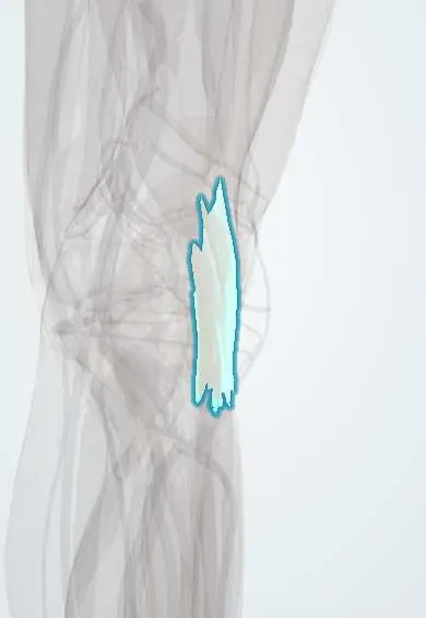
Biceps femoris tendon !
The biceps femoris tendon is the outer hamstring tendon at the back of the knee, attaching to the head of the fibula.
Official name: Tendon of biceps femoris
Opens in the Durgical app, no sign-up needed.
What it is
The outer hamstring tendon, ending on the head of the fibula.
What it does
Bends the knee and turns the shin outward; supports the outer knee.
Key facts
- Attaches
- Common tendon of both heads of biceps femoris to the head of the fibula, splitting around the fibular attachment of the LCL; slips to the lateral tibial condyle and crural fascia
- Function
- Knee flexion and lateral rotation of the leg; part of the posterolateral corner; the common fibular nerve runs along its posteromedial border
- Tight in
- Hip flexion with the knee extended
Relations
- Course
- Down the back and outer side of the thigh, forming the upper lateral border of the popliteal fossa, to the head of the fibula
- Medial
- Common fibular nerve, then the lateral head of gastrocnemius and the plantaris
- In front
- IT band and the lateral femoral condyle
- Deep
- LCL, around which its insertion splits, with a small bursa between them
How to find it
Prone or seated with the knee bent against resistance, the cord at the outer back of the knee is followed down to the fibular head; the common fibular nerve can be rolled under the fingers just behind and inside it, then around the fibular neck.
Examination
- Resisted knee flexion with the leg turned outward loads the biceps; pain or a gap near the fibular head suggests a distal tendon injury
On imaging
Knee radiographs show avulsed fragments from the fibular head; MRI shows the shared biceps–LCL attachment, tears, bone oedema and injury to the other posterolateral corner structures.
Common problems
- Strains, tendinitis, and less often a pull-off injury at the fibula with severe knee injuries.
How it is diagnosed
Physical exam; ultrasound or MRI if needed.
Treatment
- Rest and physical therapy; surgical repair for a pulled-off tendon.
Clinical notes
- Foot drop from a stretched common fibular nerve can accompany distal biceps avulsions and posterolateral corner injuries
- The tendon can snap over the fibular head, a rare cause of lateral knee snapping
Related conditions
Test yourself
Think of the answer, then tap the question to see it.
Where does the biceps femoris tendon insert, and how does it relate to the LCL?
On the head of the fibula, splitting around the fibular attachment of the LCL.
Which nerve runs along the posteromedial border of the biceps femoris tendon?
The common fibular nerve, down to the fibular neck; it is found and protected first in lateral knee surgery.
What does a bone fragment pulled off the fibular head by the biceps and LCL indicate?
A posterolateral corner injury, often with cruciate tears.
Educational information, not medical advice. For any symptom, see a health professional.
Its names
- Official Latin (TA2)
- caput longum musculi bicipitis femoris TA2 2639
- Official Spanish (DeCS)
- Tendón del bíceps femoral DeCS D013710
- English (TA2)
- Tendon of biceps femoris
Related
Sources: Terminologia Anatomica (TA2); DeCS; Anatomy, Bony Pelvis and Lower Limb, Hamstring Muscle; Anatomy, Bony Pelvis and Lower Limb: Posterior Thigh Muscles.
Spotted a mistake?We are always improving for the community. Tell us what is wrong and our team will review it.Report a mistake
Representative model, for educational purposes only.