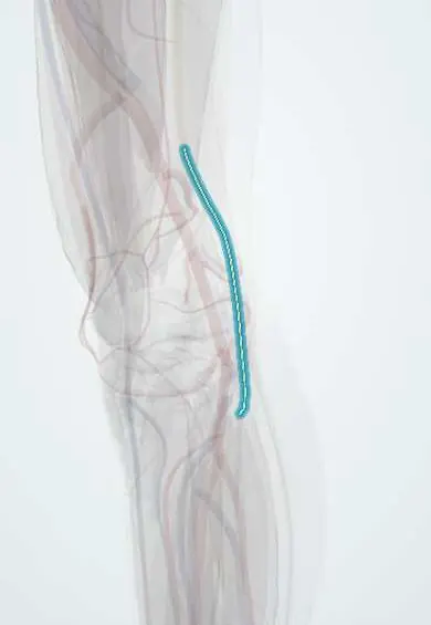
Common fibular nerve !
The common fibular nerve wraps around the outer knee just below the skin near the fibula; injury here causes foot drop.
Opens in the Durgical app, no sign-up needed.
What it is
The common peroneal (fibular) nerve, which wraps around the outer knee just below the head of the fibula.
What it does
Lifts the foot and toes, turns the foot outward, and carries feeling from the top of the foot.
Key facts
- Roots
- L4–S2
- Moves
- Short head of biceps femoris (in thigh); anterior and lateral leg via deep and superficial branches; injury at fibular neck causes foot drop
- Feeling from
- Upper lateral leg (lateral sural cutaneous nerve); dorsum of foot via terminal branches
Relations
- Course
- From the apex of the popliteal fossa it follows the medial border of the biceps femoris down the lateral side of the fossa, crosses behind the lateral head of the gastrocnemius, winds around the neck of the fibula and enters the fibularis longus, where it divides
- Lateral
- The biceps femoris tendon, whose medial edge it follows down to the head of the fibula
- Medial
- The tibial nerve and the popliteal artery and vein in the popliteal fossa
- Deep
- The lateral head of the gastrocnemius, then the bone of the fibular neck, against which it is easily pressed
- Superficial
- Only fascia and skin at the fibular neck
How to find it
With the knee slightly bent, the biceps femoris tendon stands out on the lateral side of the back of the knee; the nerve runs along its medial edge. Just below and behind the head of the fibula it can often be rolled under the fingertip as a cord crossing the fibular neck.
Examination
- Heel walking and resisted dorsiflexion of the ankle and big toe: weakness shows deep fibular involvement (foot drop)
- Resisted eversion: weakness adds superficial fibular involvement; normal inversion and hip abduction separate a fibular neck lesion from an L5 radiculopathy
- Tinel's sign at the fibular neck: tingling into the top of the foot supports compression at that point
- Sensory check on the dorsum of the foot, and in the first web space for the deep branch alone
On imaging
Ultrasound follows it along the medial edge of the biceps femoris tendon and round the fibular neck, showing swelling or a cause such as an intraneural ganglion from the proximal tibiofibular joint. Knee radiographs check for a fibular neck fracture. On MRI, denervation oedema of the anterior and lateral compartments with a normal short head of biceps places the lesion at the fibular neck.
Common problems
- Because it lies close to the skin, it can be pressed by leg crossing, a tight cast or brace, or injured in knee trauma.
- Injury can cause foot drop (difficulty lifting the foot).
How it is diagnosed
Physical exam; nerve conduction studies and EMG; imaging when needed.
Treatment
- Remove the pressure; an ankle-foot brace for foot drop; physical therapy.
- Surgery to free or repair the nerve if it does not recover.
Clinical notes
- Foot drop differential: fibular neck lesion (inversion, hip abduction and reflexes normal, no back pain) versus L5 radiculopathy (weak inversion and hip abduction, often back pain and a positive straight-leg raise); a sciatic lesion also weakens knee flexion
- At risk in fibular neck fractures, posterolateral corner and lateral collateral ligament injuries, high tibial osteotomy, lithotomy positioning, long squatting and marked weight loss
- Nerve conduction studies show slowing or block across the fibular head; EMG of the short head of the biceps femoris (normal in a fibular neck lesion, abnormal in a sciatic one) locates the level
Anatomical variants
- Fibrous bands at the origin of the fibularis longus can form a tight tunnel over the nerve at the fibular neck
- When the sciatic nerve divides high, the common fibular part may pass through or above the piriformis
Development
Built from the posterior divisions of L4–S2, it supplies muscles of the dorsal (extensor) mass of the limb bud; once the lower limb has turned medially in week 7, those muscles face forward and outward, in the anterior and lateral compartments of the leg.
Memory aid
PED and TIP: the fibular (Peroneal) nerve Everts and Dorsiflexes; the Tibial nerve Inverts and Plantarflexes. Injure PED and the foot drops.
Related conditions
- Sciatica (Lumbar Radiculopathy)
- Common Fibular (Peroneal) Nerve Injury
- Leprosy (Hansen disease)
- Charcot-Marie-Tooth disease
- Polyarteritis nodosa
- Eosinophilic granulomatosis with polyangiitis (Churg–Strauss)
Test yourself
Think of the answer, then tap the question to see it.
Why is the common fibular nerve the most often injured nerve of the lower limb?
It winds around the neck of the fibula with only skin over it, so it is pressed by leg crossing, tight casts, fractures and knee trauma.
What deficits follow a common fibular nerve lesion at the fibular neck?
Foot drop with weak eversion, numbness on the dorsum of the foot, but normal inversion and a normal Achilles reflex.
How is a fibular neck lesion told apart from an L5 radiculopathy?
In L5 radiculopathy inversion and hip abduction are also weak, often with back pain and a positive straight-leg raise; a fibular neck lesion spares them.
Where does the common fibular nerve divide, and into what?
At the fibular neck, inside the fibularis longus, into the superficial and deep fibular nerves.
What does the mnemonic PED and TIP mean?
The fibular (Peroneal) nerve Everts and Dorsiflexes; the Tibial nerve Inverts and Plantarflexes. Injure PED and the foot drops.
Educational information, not medical advice. For any symptom, see a health professional.
Its names
- Official Latin (TA2)
- nervus fibularis communis TA2 6571
- Official Spanish (DeCS)
- Nervio peroneo DeCS D010543
- English (TA2)
- Common fibular nerve
- Also called
- common peroneal nerve · peroneal nerve · nervus peronaeus commune
Related
- Sciatic nerve
- Biceps femoris tendon
- Deep fibular nerve
- Superficial fibular nerve
- Tibialis anterior
- Extensor digitorum longus
- Fibularis longus
- Fibularis brevis
Sources: Terminologia Anatomica (TA2); DeCS; Anatomy, Bony Pelvis and Lower Limb: Calf Common Peroneal Nerve (Common Fibular Nerve); 8.5 Development of the Appendicular Skeleton (OpenStax (Rice University), 2022) · adapted, CC BY 4.0; Anatomía humana (4.ª ed.), tomo 1, cap. 70: Nervios del plexo lumbosacro.
Spotted a mistake?We are always improving for the community. Tell us what is wrong and our team will review it.Report a mistake
Representative model, for educational purposes only.