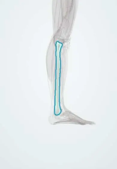
Fibula !
The fibula is the thin bone on the outer side of the lower leg; its lower end forms the bony bump on the outside of the ankle.
Opens in the Durgical app, no sign-up needed.
What it is
The thin outer bone of the leg, from just below the knee to the ankle.
What it does
Its head anchors the LCL. Its lower end forms the outer ankle bone (lateral malleolus), which helps hold the talus in place. It carries little weight.
Key facts
- Joints with
- Tibia (superior and inferior tibiofibular joints); talus (ankle); not part of the knee joint
- Landmarks
- Head with apex; neck (common fibular nerve winds around it); lateral malleolus; malleolar fossa
Relations
- Above
- Head: attachment of the lateral collateral ligament and the biceps femoris tendon; the common fibular nerve runs behind the tendon, then crosses the neck and divides into the superficial and deep fibular nerves
- Lateral
- Fibularis longus and fibularis brevis on the lateral surface; their tendons share a groove behind the lateral malleolus under the superior fibular retinaculum
- In front
- Extensor digitorum longus, extensor hallucis longus and fibularis tertius arise here; the anterior tibial artery and deep fibular nerve lie nearby on the interosseous membrane
- Behind
- Soleus and flexor hallucis longus; the fibular artery runs close to the back of the bone
- Medial
- Interosseous membrane joining it to the tibia; tibialis posterior arises from both bones and the membrane
How to find it
The head is felt on the posterolateral side of the knee at about the level of the tibial tuberosity, by following the biceps femoris tendon down. The common fibular nerve can be rolled against the neck just below and behind it. The shaft is buried in muscle until its lower quarter, then ends as the subcutaneous lateral malleolus, which reaches lower and lies further back than the medial malleolus.
Examination
- Ottawa ankle rules: tenderness at the posterior edge or tip of the lateral malleolus (lower 6 cm), or inability to bear weight, indicates an ankle X-ray
- Squeeze test: compressing the tibia and fibula together at mid-calf causes pain at the ankle in a syndesmotic (high ankle) sprain
On imaging
Ankle series (AP, mortise, lateral): the fracture level gives the Weber type; a widened medial clear space or reduced tibiofibular overlap points to syndesmotic injury. An ankle injury with medial tenderness and no lateral malleolus fracture warrants views of the whole leg to find a proximal (Maisonneuve) fracture.
Common problems
- Fractures of the outer ankle bone are among the most common ankle fractures.
- Stress fractures in runners.
Clinical notes
- After a fibular neck fracture or a lateral knee injury, ankle dorsiflexion and sensation on the dorsum of the foot are checked to assess the common fibular nerve
- Free fibula flap for jaw and long-bone reconstruction; a distal segment is left in place to keep the ankle stable
- A lateral malleolus fracture with a shifted talus makes the ankle mortise unstable
Development
The distal centre appears before the proximal one but also fuses first: the classic exception to the rule that the epiphysis that appears first fuses last.
Related conditions
Test yourself
Think of the answer, then tap the question to see it.
Which nerve winds around the fibular neck, and what deficit follows its injury there?
The common fibular nerve; fractures, tight casts or prolonged pressure there cause foot drop.
How does the Weber classification grade lateral malleolus fractures?
By level relative to the syndesmosis: A below, B at, C above. Type C means the syndesmosis is torn.
What is a Maisonneuve fracture?
A proximal fibular fracture with a medial ankle injury and a torn syndesmosis; hence the whole fibula is palpated in ankle injuries.
What does the squeeze test detect at the ankle?
A syndesmotic (high ankle) sprain: compressing tibia and fibula together at mid-calf causes pain at the ankle.
Why can the middle part of the fibula be used as a free vascularised bone graft?
It carries little weight; the graft is taken on the fibular artery, leaving a distal segment to keep the ankle stable.
Educational information, not medical advice. For any symptom, see a health professional.
Its names
- Official Latin (TA2)
- fibula TA2 1427
- Official Spanish (DeCS)
- Peroné DeCS D005360
- English (TA2)
- Fibula
- Also called
- os fibulare · fibular bone
Related
- Arcuate popliteal ligament
- ATFL
- Biceps femoris (long head)
- Biceps femoris tendon
- Biceps femoris (short head)
- Biceps femoris tendon (short head)
- CFL
- Extensor digitorum longus
Sources: Terminologia Anatomica (TA2); DeCS; Anatomy, Bony Pelvis and Lower Limb: Fibula; Anatomy, Bony Pelvis and Lower Limb: Leg Bones.
Spotted a mistake?We are always improving for the community. Tell us what is wrong and our team will review it.Report a mistake
Representative model, for educational purposes only.