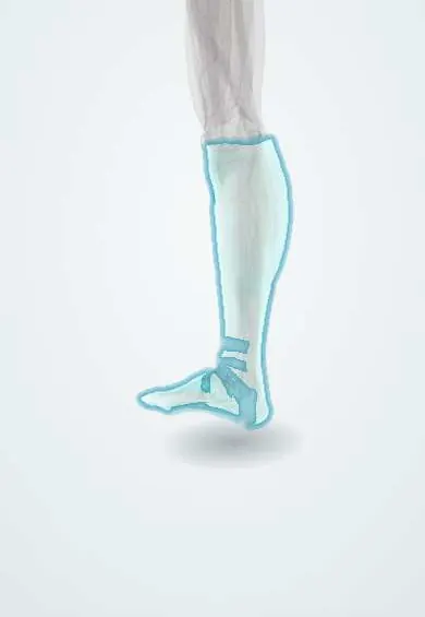
Crural fascia !
The crural fascia is a thin, tough sheet wrapping the leg muscles from knee to ankle; it divides the leg into closed compartments.
Opens in the Durgical app, no sign-up needed.
What it is
A thin, tough sheet wrapping the leg muscles from the knee to the ankle.
What it does
Holds the leg muscles in their compartments. Near the ankle it thickens into the retinacula that hold the tendons in place.
Key facts
- Where
- Deep fascia of leg from knee to ankle; continuous with fascia lata above
- Attaches
- Anterior and medial borders of tibia, fibular head and malleoli; thickens into ankle retinacula
- Function
- With intermuscular septa forms tight anterior, lateral and posterior compartments (compartment syndrome)
Relations
- Superficial
- Subcutaneous great and small saphenous veins, the saphenous and sural nerves; the superficial fibular nerve pierces it in the lower third of the lateral leg
- Deep
- Blends with the periosteum of the subcutaneous medial surface of the tibia and sends the anterior and posterior intermuscular septa to the fibula
- Above
- Continuous with the fascia lata and the popliteal fascia; receives expansions from the sartorius, gracilis, semitendinosus and biceps femoris tendons
- Below
- Thickens into the extensor, flexor and fibular retinacula at the ankle
- Contents
- Anterior: tibialis anterior, toe extensors, fibularis tertius, anterior tibial artery, deep fibular nerve. Lateral: fibularis longus and brevis, superficial fibular nerve. Superficial posterior: gastrocnemius, soleus, plantaris. Deep posterior: tibialis posterior, long toe flexors, popliteus, posterior tibial and fibular vessels, tibial nerve
How to find it
It is not felt as a separate layer; the tension of each compartment is judged by squeezing it. The anterolateral fasciotomy incision is described roughly midway between the tibial crest and the fibula, and the posteromedial one a little behind the medial border of the tibia.
Examination
- Pain on passive stretch of a compartment's muscles (for example, passive toe flexion for the anterior compartment) is an early clinical sign of compartment syndrome
- Compartment pressure measurement: a needle manometer in each compartment; a small gap between diastolic blood pressure and compartment pressure (commonly under 30 mmHg) supports the diagnosis
On imaging
Imaging has a small role in acute compartment syndrome. MRI shows muscle oedema limited to one compartment; dynamic ultrasound shows defects in the fascia with muscle bulging through them on contraction.
Common problems
- Compartment syndrome: swelling inside a tight compartment squeezes the muscles and nerves. After an injury it is an emergency; in some athletes it comes on with exercise.
Clinical notes
- In the anterolateral incision the anterior intermuscular septum is found first and the fascia is opened in front of it (anterior compartment) and behind it (lateral compartment)
- The deep posterior compartment is the one most often missed; it is reached from the posteromedial incision by detaching soleus from the tibia
- The great saphenous vein and saphenous nerve lie along the posteromedial incision and are kept out of harm
- Fascial defects with muscle hernia are most often found where the superficial fibular nerve exits, and can cause local pain or nerve symptoms
Related conditions
- Acute compartment syndrome
- Cellulitis of the leg
- Lymphoedema
- Medial tibial stress syndrome (shin splints)
Test yourself
Think of the answer, then tap the question to see it.
Which four compartments of the leg does the crural fascia close, with the septa and interosseous membrane?
Anterior, lateral, superficial posterior and deep posterior; the anterior compartment is the one most often affected by compartment syndrome.
What early sensory sign suggests anterior compartment syndrome of the leg?
Numbness in the first web space of the foot, the territory of the deep fibular nerve, which runs in the anterior compartment.
Which two incisions are used for a standard leg fasciotomy through the crural fascia?
An anterolateral incision for the anterior and lateral compartments and a posteromedial incision for the superficial and deep posterior compartments.
Which nerve pierces the crural fascia in the lower lateral leg and is at risk in the anterolateral fasciotomy incision?
The superficial fibular nerve; fascial defects with muscle hernia are also most often found where it exits.
Educational information, not medical advice. For any symptom, see a health professional.
Its names
- Official Latin (TA2)
- fascia cruris TA2 2708
- Official Spanish (DeCS)
- Fascia de la pierna DeCS D005205
- English (TA2)
- Crural fascia
- Also called
- deep fascia of leg
Related
Sources: Terminologia Anatomica (TA2); DeCS; Anatomy, Fascia; Anatomy, Bony Pelvis and Lower Limb: Leg Anterior Compartment.
Spotted a mistake?We are always improving for the community. Tell us what is wrong and our team will review it.Report a mistake
Representative model, for educational purposes only.