Joints and ligaments · Lower limb
LCL !
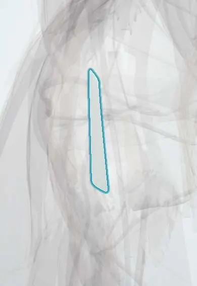
The lateral collateral ligament (LCL) is a cord on the outer side of the knee, from femur to fibula; it keeps the knee from bending outward.
Official name: Fibular collateral ligament
Opens in the Durgical app, no sign-up needed.
What it is
A cord-like band on the outer side of the knee, from the femur to the head of the fibula.
What it does
Keeps the knee from bending outward.
Key facts
- Attaches
- Lateral epicondyle of femur to lateral surface of fibular head; separate from capsule and lateral meniscus
- Function
- Resists varus stress; limits external rotation of tibia; part of posterolateral corner
- Tight in
- Knee extension; varus stress
Relations
- Superficial
- Biceps femoris tendon, which splits around the LCL as both insert on the fibular head
- Deep
- Popliteus tendon, passing between it and the lateral meniscus; the inferior lateral genicular artery runs between it and the capsule
- Behind
- Common fibular nerve, behind the biceps tendon, heading for the fibular neck
- In front
- IT band (iliotibial band) ending on Gerdy tubercle, and the anterolateral capsule
How to find it
Seated with the leg crossed in a figure-of-four (ankle resting on the opposite knee), the ligament stands out as a firm, pencil-thick cord between the lateral femoral epicondyle and the fibular head, just in front of the biceps tendon.
Examination
- Varus stress at 30°: lateral opening compared with the other knee means an LCL tear
- Varus stress at 0°: opening in full extension adds posterolateral corner and often cruciate injury
- Dial test: over 10° more external rotation than the other side at 30° points to the posterolateral corner
- External rotation recurvatum test: lifted by the big toes, the injured knee falls into hyperextension, varus and external rotation (posterolateral corner, often with a cruciate)
On imaging
MRI: a thin dark band from the lateral epicondyle to the fibular head, seen across several posterior coronal slices because it runs obliquely. On X-ray, a small avulsion of the fibular head apex (arcuate sign) marks a posterolateral corner injury.
Common problems
- Less often injured alone; usually from a blow to the inner side of the knee.
- Often part of an injury to the back-outer corner of the knee.
How it is diagnosed
An exam test that gently pushes the knee outward (varus stress test). MRI.
Treatment
- Mild injuries: brace and physical therapy.
- Severe or combined injuries: surgical repair or reconstruction.
Clinical notes
- An unrecognised posterolateral corner injury is a classic cause of ACL or PCL graft failure
- Foot drop after a varus knee injury suggests common fibular nerve damage; ankle dorsiflexion and sensation on the dorsum of the foot are checked
- Anatomic posterolateral corner reconstruction rebuilds the LCL, popliteus tendon and popliteofibular ligament with grafts passed through the fibular head and lateral femur
Test yourself
Think of the answer, then tap the question to see it.
What are the attachments of the lateral collateral ligament (LCL)?
Lateral epicondyle of the femur to the lateral surface of the fibular head; it is separate from the capsule and the lateral meniscus.
At what knee position does varus stress isolate the LCL?
At 30° of flexion. Opening also in full extension points to cruciate and posterolateral corner damage.
What are the main static stabilizers of the posterolateral corner of the knee?
The LCL, the popliteus tendon and the popliteofibular ligament.
What does foot drop after a varus knee injury with an LCL tear suggest?
Common fibular nerve damage: the nerve winds round the fibular neck just below the LCL insertion.
Where is the LCL felt on examination?
In the figure-of-four position, as a firm, pencil-thick cord between the lateral femoral epicondyle and the fibular head, in front of the biceps tendon.
Educational information, not medical advice. For any symptom, see a health professional.
Its names
- Official Latin (TA2)
- ligamentum collaterale fibulare TA2 1895
- Official Spanish (DeCS)
- Ligamento colateral fibular (peroneo) DeCS D017885
- English (TA2)
- Fibular collateral ligament
- Also called
- lateral collateral ligament of knee
Related
- Biceps femoris tendon
- Popliteus tendon
- Popliteofibular ligament
- IT band
- Anterolateral ligament (ALL)
- Femur
- Fibula
Sources: Terminologia Anatomica (TA2); DeCS; Anatomy, Bony Pelvis and Lower Limb, Knee.
Spotted a mistake?We are always improving for the community. Tell us what is wrong and our team will review it.Report a mistake
Representative model, for educational purposes only.