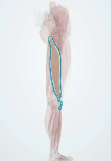
Biceps femoris (long head) !
The long head of biceps femoris is the outer hamstring muscle, from the sitting bone; it bends the knee, extends the hip and is often strained.
Official name: Long head of biceps femoris
Opens in the Durgical app, no sign-up needed.
What it is
The outer hamstring muscle at the back of the thigh.
What it does
Bends the knee, extends the hip and turns the shin outward.
Key facts
- Origin
- Ischial tuberosity (common tendon with semitendinosus); sacrotuberous ligament
- Insertion
- Lateral side of head of fibula (tendon split by lateral collateral ligament); slips to the lateral tibial condyle and crural fascia
- Action
- Extends hip; flexes knee; laterally rotates flexed knee
- Nerve
- Sciatic nerve, tibial division (L5–S2)
- Blood supply
- Perforating arteries; inferior gluteal artery; medial circumflex femoral artery
Relations
- Course
- From the ischial tuberosity it runs down and laterally across the back of the thigh, joins the short head in the lower third and ends on the head of the fibula
- Superficial
- Gluteus maximus over its origin, then fascia lata and skin
- Deep
- Sciatic nerve, which it crosses, and adductor magnus; lower down, the short head
- Medial
- Semitendinosus, which shares its origin; the two diverge above the knee, the biceps forming the upper lateral border of the popliteal fossa
- Below
- Lateral collateral ligament, which splits its tendon; the common fibular nerve runs along the posteromedial edge of the tendon and winds around the fibular neck
How to find it
Prone with the knee bent against resistance and the leg turned slightly outward, the thick tendon stands out at the outer side of the back of the knee and can be followed to the head of the fibula; the common fibular nerve can often be felt as a cord just behind it.
Examination
- Passive straight-leg raise and popliteal angle: posterior thigh pain or a smaller range on the injured side points to hamstring strain or tightness; pain spreading below the knee suggests nerve root tension instead
- Resisted knee flexion with the leg turned outward emphasizes the biceps femoris; pain or weakness suggests a lateral hamstring injury
On imaging
MRI is the main test for hamstring strains: fluid-sensitive images show feathery oedema around its proximal muscle–tendon junction, the classic site; a gap or tendon involvement means a more severe tear. Ultrasound is a quick option for acute strains. At the ischial tuberosity, MRI shows avulsion of the conjoint tendon, and on axial images the sciatic nerve lies just deep to the muscle.
Common problems
- Hamstring strain, common in sprinting sports.
- Rarely, the tendon pulls off the bone at the hip.
How it is diagnosed
Physical exam; MRI for severe strains.
Treatment
- Rest, then progressive stretching and strengthening; surgery only for complete tendon pull-offs.
Clinical notes
- Proximal avulsion of the conjoint tendon (with the semitendinosus): bruising of the back of the thigh and a palpable gap; complete tears of several tendons with retraction are often repaired, and the nearby sciatic nerve can be caught in scar in late cases
- Avulsion fracture of the fibular head (arcuate sign) at the attachment of the biceps femoris and the lateral collateral ligament signals a posterolateral corner injury
- Operations at the lateral knee, such as posterolateral corner reconstruction, put the common fibular nerve at risk along the medial edge of the tendon
Development
The long head comes from the ventral (flexor) muscle mass and keeps the tibial-division supply of the hamstrings; the short head has a different origin and nerve, so the two heads are joined only at the tendon.
Related conditions
Test yourself
Think of the answer, then tap the question to see it.
Which nerve supplies the long head of the biceps femoris, and how does it differ from the short head?
The tibial division of the sciatic nerve; the short head is supplied by the common fibular division.
Which hamstring is most often strained, and where?
The long head of the biceps femoris, usually at the proximal muscle–tendon junction during high-speed running.
Which nerve follows the medial edge of the biceps femoris tendon to the fibular neck?
The common fibular nerve; it is at risk along this edge in lateral knee surgery such as posterolateral corner reconstruction.
What does an avulsion fracture of the fibular head (arcuate sign) indicate?
A posterolateral corner injury, at the attachment of the biceps femoris and the lateral collateral ligament.
Educational information, not medical advice. For any symptom, see a health professional.
Its names
- Official Latin (TA2)
- caput longum musculi bicipitis femoris TA2 2639
- Official Spanish (DeCS)
- Cabeza larga del músculo bíceps femoral DeCS D000070633
- English (TA2)
- Long head of biceps femoris
Related
- Biceps femoris (short head)
- Sacrotuberous ligament
- Sciatic nerve
- Perforating arteries
- Biceps femoris tendon
- Fibula
Sources: Terminologia Anatomica (TA2); DeCS; Anatomy, Bony Pelvis and Lower Limb, Hamstring Muscle; Anatomy, Bony Pelvis and Lower Limb: Posterior Thigh Muscles.
Spotted a mistake?We are always improving for the community. Tell us what is wrong and our team will review it.Report a mistake
Representative model, for educational purposes only.