Muscular system · Head and neck
Arytenoid muscles !
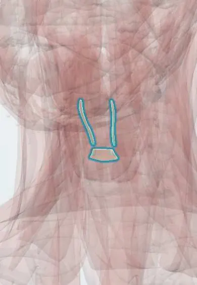
The arytenoid muscles are small muscles between the two arytenoid cartilages at the back of the larynx; they close the back of the vocal folds.
Official name: Transverse arytenoid muscle
Opens in the Durgical app, no sign-up needed.
What it is
Small muscles between the two arytenoids.
What it does
Close the back of the vocal folds.
Key facts
- Origin
- One arytenoid
- Insertion
- The opposite arytenoid (oblique part continues as aryepiglottic)
- Action
- Close the back of the glottis
- Nerve
- Recurrent laryngeal nerve (X)
- Blood supply
- Laryngeal arteries
Relations
- Behind
- Mucosa of the front wall of the laryngopharynx, above the cricoid lamina
- Lateral
- Posterior surfaces of both arytenoid cartilages, which they bridge
- Below
- Posterior cricoarytenoid muscles on the back of the cricoid lamina
- Above
- Aryepiglottic folds, which the oblique fibres enter on their way to the epiglottis
How to find it
Not palpable. On laryngoscopy their area is the interarytenoid mucosa between the two arytenoid mounds, which narrows as the arytenoids come together on phonation.
Examination
- Laryngoscopy on a sustained vowel: a gap left at the back of the glottis suggests weak posterior closure, although a small posterior gap is common in healthy voices, especially in women
On imaging
Not assessed on their own: the transverse and oblique arytenoids lie in the midline on the back of the arytenoid cartilages, just in front of the hypopharynx. Laryngoscopy and stroboscopy judge closure of the back of the glottis. Contrast CT or MRI check this posterior commissure for tumour spread, which changes staging, and CT shows the arytenoids when dislocation after intubation is suspected.
Clinical notes
- The interarytenoid region is where reflux of stomach contents into the larynx often shows: redness and thickened mucosa (posterior laryngitis)
- Posterior glottic stenosis after prolonged intubation scars this region, tethers the arytenoids and limits opening, mimicking bilateral paralysis
- Swallowing manoeuvres used in dysphagia therapy, such as the supraglottic swallow (holding the breath before and during the swallow), rely on firm closure of the arytenoids and the laryngeal inlet
Development
From sixth-arch mesoderm, hence the recurrent laryngeal nerve. If the back of the larynx fails to close in the midline, a posterior laryngeal cleft opens between the arytenoids, causing choking and aspiration in infants.
Test yourself
Think of the answer, then tap the question to see it.
Which is the only unpaired intrinsic laryngeal muscle?
The transverse arytenoid; it pulls the arytenoid cartilages together and closes the back of the glottis.
Where do the oblique arytenoid fibres continue, and what do they do in swallowing?
They cross in an X and continue as the aryepiglottic muscles, narrowing the laryngeal inlet like a purse string.
What does laryngopharyngeal reflux often show in the interarytenoid region?
Redness and thickened mucosa (posterior laryngitis).
Educational information, not medical advice. For any symptom, see a health professional.
Its names
- Official Latin (TA2)
- musculus arytenoideus transversus TA2 2205
- Official Spanish (DeCS)
- Músculos aritenoideos DeCS D007821
- English (TA2)
- Transverse arytenoid muscle
- Also called
- pars transversa musculi arytaenoidei
Related
Sources: Terminologia Anatomica (TA2); DeCS; Anatomy, Head and Neck: Laryngeal Muscles; Anatomy, Head and Neck: Larynx Muscles.
Spotted a mistake?We are always improving for the community. Tell us what is wrong and our team will review it.Report a mistake
Representative model, for educational purposes only.