Muscular system · Head and neck
Posterior cricoarytenoid !
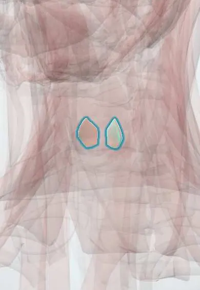
The posterior cricoarytenoid is a muscle on the back of the larynx; it is the only muscle that opens the vocal folds to let air in.
Official name: Musculus cricoarytenoideus posterior
Opens in the Durgical app, no sign-up needed.
What it is
A muscle on the back of the cricoid.
What it does
The only muscle that opens the vocal folds for breathing.
Key facts
- Origin
- Posterior cricoid lamina
- Insertion
- Muscular process of the arytenoid
- Action
- Abducts the vocal folds (the only opener)
- Nerve
- Recurrent laryngeal nerve (X)
- Blood supply
- Inferior laryngeal artery
Relations
- Behind
- Mucosa of the front wall of the laryngopharynx; just below, the start of the oesophagus
- Above
- Transverse and oblique arytenoid muscles between the two arytenoids
- Lateral
- Recurrent laryngeal nerve and inferior laryngeal artery entering the larynx near the cricothyroid joint; the lateral cricoarytenoid in front of the muscular process
- Medial
- Its partner of the other side, separated by the vertical ridge of the cricoid lamina
How to find it
Not palpable. On laryngoscopy its action is the folds swinging apart on a sniff or deep breath in. In dissection it is found by turning the larynx over and removing the laryngopharynx mucosa behind the cricoid cartilage.
Examination
- Laryngoscopy on sniffing: the folds should open; a fold that stays put points to nerve palsy, a fixed cricoarytenoid joint or posterior glottic scar, and laryngeal electromyography helps separate them
On imaging
Contrast CT or MRI of the neck shows it on axial images as a thin muscle on the back of the cricoid lamina, facing the hypopharynx. In recurrent laryngeal nerve palsy it shrinks and turns fatty; with a widened piriform sinus and a fold sitting near the midline on the same side, this is the classic CT picture. Imaging then follows the nerve down to find the cause, on the left into the chest.
Clinical notes
- Bilateral palsy most often follows thyroid surgery, neck or chest tumours or prolonged intubation; treatment ranges from tracheostomy to widening the posterior glottis (cordotomy, arytenoidectomy)
- The left recurrent laryngeal nerve hooks under the aortic arch, so chest causes (lung or oesophageal cancer, mediastinal nodes, aortic aneurysm) affect the left side more often
- Nocturnal stridor in multiple system atrophy reflects poor vocal fold opening during sleep and is linked to sudden death in that disease
Development
Like every intrinsic laryngeal muscle except the cricothyroid, it develops from sixth-arch mesoderm, which explains its supply by the recurrent laryngeal nerve.
Memory aid
"Posterior cricoarytenoid Pulls Cords Apart": the only opener; all the others close or tense the glottis.
Test yourself
Think of the answer, then tap the question to see it.
Which is the only muscle that abducts the vocal folds?
The posterior cricoarytenoid; it rotates the arytenoid cartilages outward and opens the glottis with each breath in.
What are the origin, insertion and nerve of the posterior cricoarytenoid?
From the posterior cricoid lamina to the muscular process of the arytenoid; supplied by the recurrent laryngeal nerve (X).
Why can bilateral recurrent laryngeal nerve palsy need an urgent airway?
Both posterior cricoarytenoids are lost, so the folds stay near the midline: the voice may be fair but breathing is blocked (stridor).
Why do chest diseases affect the left recurrent laryngeal nerve more often?
The left recurrent laryngeal nerve hooks under the aortic arch, near lung or oesophageal cancer, mediastinal nodes and aortic aneurysm.
Educational information, not medical advice. For any symptom, see a health professional.
Its names
- Official Latin (TA2)
- musculus cricoarytenoideus posterior TA2 2196
- Official Spanish (DeCS)
- Músculo cricoaritenoideo posterior DeCS D007821
- English (TA2)
- Musculus cricoarytenoideus posterior
- Also called
- posticus · musculus cricoarytaenoideus dorsalis
Related
Sources: Terminologia Anatomica (TA2); DeCS; Anatomy, Head and Neck: Cervical, Respiratory, Larynx, and Cricoarytenoid; Anatomy, Head and Neck: Laryngeal Muscles.
Spotted a mistake?We are always improving for the community. Tell us what is wrong and our team will review it.Report a mistake
Representative model, for educational purposes only.