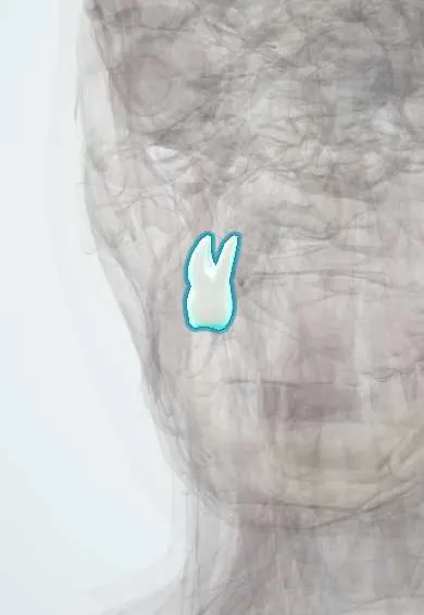
Digestive system · Head and neck
Upper right second molar !
The upper right second molar (tooth 17) is the back tooth behind the first molar that comes in around age 12; it grinds food.
Opens in the Durgical app, no sign-up needed.
What it is
A large back tooth behind the first molar, with three roots.
What it does
Grinds food before swallowing.
Key facts
- Joints with
- Alveolar socket of the maxilla (gomphosis held by the periodontal ligament); bites against the opposing teeth
- Landmarks
- FDI 17, upper right second molar: 3 roots; erupts behind the first molar around age 12; erupts at about 12–13 years; nerve: posterior superior alveolar nerve (V2); roots near the floor of the maxillary sinus
Relations
- In front
- The upper right first molar
- Behind
- The upper right third molar
- Lateral
- Cheek and buccinator; the parotid duct opens on the inner cheek opposite this tooth, at a small papilla
- Above
- Floor of the maxillary sinus
- Below
- Opposes the lower right second molar and the lower right third molar
How to find it
Seventh tooth from the midline on the upper right; the small papilla of the parotid duct on the cheek opposite its crown is a handy landmark.
On imaging
On a panoramic film its close-set roots often overlap the sinus floor; periapicals and bitewings show its roots and contacts, and CBCT clarifies a root that seems to sit in the sinus.
Common problems
- Tooth decay (caries) in the grooves of the chewing surface.
Clinical notes
- Posterior superior alveolar nerve: it enters the back of the maxilla at the tuberosity, where the posterior superior alveolar block is placed; the pterygoid venous plexus lies close by, so a haematoma can follow that injection
- Extraction can open the maxillary sinus (oroantral communication) because its roots and the sinus floor are so close
Development
No primary predecessor: it develops in the maxillary tuberosity behind the first molar and erupts once the jaw has grown enough to make room.
Related conditions
Test yourself
Think of the answer, then tap the question to see it.
Where is the posterior superior alveolar block given for the upper right second molar, and what is its risk?
At the maxillary tuberosity, where the nerve enters the maxilla; the nearby pterygoid venous plexus can bleed, giving a haematoma.
How do the roots of the upper right second molar differ from the first molar's?
They sit closer together, may be fused, and are often the roots nearest to the maxillary sinus floor.
Which primary tooth does the upper right second molar replace?
None: it develops in the maxillary tuberosity behind the first molar.
At about what age does the upper right second molar erupt?
At about 12 years, behind the first molar, with no primary tooth lost to make room.
Educational information, not medical advice. For any symptom, see a health professional.
Related
Sources: Anatomy, Head and Neck, Teeth; Anatomy, Permanent Dentition; Anatomy, Head and Neck, Tooth Eruption; Anatomy, Head and Neck: Alveolar Nerve; 23.3 The Mouth, Pharynx, and Esophagus (OpenStax (Rice University), 2022) · adapted, CC BY 4.0.
Spotted a mistake?We are always improving for the community. Tell us what is wrong and our team will review it.Report a mistake
Representative model, for educational purposes only.