Respiratory system · Head and neck
Maxillary sinus !
The maxillary sinus is the largest air space of the face, in the upper jawbone under each eye; it drains into the nose, just above the upper molar roots.
Opens in the Durgical app, no sign-up needed.
What it is
The largest air space, inside each maxilla under the eye.
What it does
Drains into the nose; upper molar roots sit just below it.
Key facts
- Where
- Body of the maxilla; opens high into the middle meatus
- Blood supply
- Maxillary artery branches
- Nerve
- V2 (superior alveolar, infraorbital nerves)
- Function
- Lightens the skull, resonance; drains poorly (ostium high)
Relations
- Above
- Orbital floor, with the infraorbital nerve and vessels in their groove and canal
- Below
- Alveolar process with the roots of the upper premolars and molars; in adults the floor lies below the level of the nasal floor
- Medial
- Lateral wall of the nasal cavity: inferior meatus, middle meatus, uncinate process and the natural ostium in the ethmoidal infundibulum
- Behind
- Pterygopalatine and infratemporal fossae, with the maxillary artery and posterior superior alveolar nerves
- In front
- Facial surface of the maxilla, the infraorbital foramen and the soft tissues of the cheek
How to find it
It cannot be felt; tenderness over the cheek and upper teeth pain that worsens on bending forward can accompany sinusitis. Its front wall is reached through the mouth above the canine and premolar roots (canine fossa).
Examination
- Transillumination with a light inside the mouth in a dark room: a dull glow on one side suggests fluid or thick lining, but the sign is unreliable and has been largely replaced by endoscopy and CT
On imaging
Sinus CT shows mucosal thickening, fluid levels, ostiomeatal complex blockage and the relation of the tooth roots to the floor; dental imaging (panoramic, cone-beam CT) shows root apices and oroantral fistulas. Blood in the sinus after facial trauma suggests a fracture.
Common problems
- Sinusitis: blocked nose, thick discharge, pressure in the cheeks or teeth.
- Most acute sinusitis is viral; some becomes chronic.
How it is diagnosed
Symptoms and exam; nasal endoscopy and CT for chronic or complicated cases.
Treatment
- Saline rinses, nasal steroid sprays, pain relief; antibiotics only for bacterial cases.
- Endoscopic sinus surgery for chronic sinusitis.
Clinical notes
- An oroantral communication after an upper molar extraction lets fluid pass from mouth to nose; small ones heal on their own, larger ones may need a flap
- A sinus lift (floor augmentation) raises the sinus lining to make room for dental implants in the back of the upper jaw
- The Caldwell–Luc approach opened the sinus through the canine fossa; endoscopic middle meatal antrostomy has largely replaced it
Anatomical variants
- Bony septa (Underwood septa) that divide the floor into recesses, relevant to sinus lifts and implants
- Accessory ostia in the middle meatus, which can let mucus circulate back into the sinus
Development
Present at birth as a small cavity; it grows in two spurts, in early childhood and with eruption of the permanent teeth, and its floor reaches the level of the nasal floor around early adolescence.
Related conditions
Test yourself
Think of the answer, then tap the question to see it.
Why can maxillary sinusitis feel like upper toothache?
The walls of the maxillary sinus carry the superior alveolar nerves, and the upper molar and premolar roots lie close to its floor.
What dental problems can involve the maxillary sinus?
Dental infection can cause odontogenic sinusitis, and an upper molar extraction can open an oroantral communication.
What does one-sided maxillary sinus disease with bone erosion or bleeding suggest?
A tumour or a dental cause rather than simple sinusitis.
Why does the maxillary sinus drain poorly?
Its ostium opens high into the middle meatus.
Educational information, not medical advice. For any symptom, see a health professional.
Its names
- Official Latin (TA2)
- sinus maxillaris TA2 3177
- Official Spanish (DeCS)
- Seno Maxilar DeCS D008443
- English (TA2)
- Maxillary sinus
- Also called
- antrum of Highmore · maxillary paranasal sinus · sinus Highmori
Related
Sources: Terminologia Anatomica (TA2); DeCS; Anatomy, Head and Neck, Nose Paranasal Sinuses; Anatomy, Head and Neck, Nose Sinuses.
Spotted a mistake?We are always improving for the community. Tell us what is wrong and our team will review it.Report a mistake
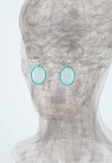
Representative model, for educational purposes only.