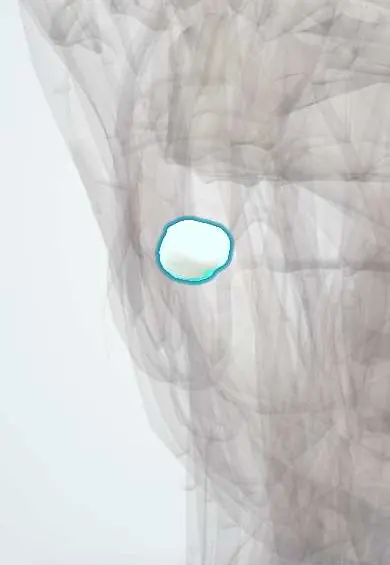
Digestive system · Head and neck
Lower right third molar !
The lower right third molar (tooth 48) is the wisdom tooth, last at the back; it often lacks room to come in and stays stuck (impacted).
Opens in the Durgical app, no sign-up needed.
What it is
The wisdom tooth: the last molar, usually coming in between the ages of 17 and 25.
What it does
Grinds food when it erupts fully; often it does not.
Key facts
- Joints with
- Alveolar socket of the mandible (gomphosis held by the periodontal ligament); bites against the opposing teeth
- Landmarks
- FDI 48, lower right third molar (wisdom tooth): roots often fused or variable; often impacted or missing; erupts at about 17–25 years; nerve: inferior alveolar nerve (V3); close to the inferior alveolar canal and the lingual nerve
Relations
- In front
- The lower right second molar
- Behind
- Ramus of the mandible; Pell and Gregory's classification grades how much room the ramus leaves and how deep the tooth lies
- Above
- Meets the upper right second molar and the upper right third molar, when erupted
- Below
- Inferior alveolar canal and nerve, often in contact with the roots
- Medial
- Lingual nerve, just inside the lingual plate at this level
How to find it
Eighth and last tooth on the lower right, in front of the ramus; often hidden in bone or partly covered by a flap of gum behind the second molar.
On imaging
A panoramic film comes first. Darkening of the root where the canal crosses, an interrupted white canal outline or a canal that bends around the root suggest close contact; CBCT then maps it.
Common problems
- Impacted wisdom tooth: stuck under the gum or against the second molar, with pain, swelling or infection (pericoronitis).
Clinical notes
- Inferior alveolar nerve block for anaesthesia; the buccal nerve covers the cheek-side gum around and behind it
- A partly erupted crown under a flap of gum (operculum) collects food and bacteria, the setting of pericoronitis; spread to the masticator spaces limits mouth opening
- When the roots are closely tied to the canal, coronectomy (removing only the crown) can spare the inferior alveolar nerve
- A dentigerous cyst can develop around the crown while it stays unerupted
Anatomical variants
- Its shape is very variable: cusps may be added or lost, and the roots are often fused into one cone or sharply curved
Development
Its germ starts to calcify at about 9–10 years and the roots finish in the early twenties, the basis of forensic age estimates; one or more third molars are often missing.
Memory aid
FDI 48 = quadrant 4 (lower right), tooth 8. In the US Universal system it is #32, the last number of the count.
Related conditions
Test yourself
Think of the answer, then tap the question to see it.
Which is the most often impacted tooth, and what is its most common tilt?
The lower third molar, such as the lower right third molar; mesioangular is the most common tilt in Winter's classification.
What deficit follows injury to the inferior alveolar nerve during removal of the lower right third molar?
Numbness of the lower lip and chin on that side, because the roots often lie close to or across the inferior alveolar canal.
What is pericoronitis of the lower right third molar, and what sign shows spread to the masticator spaces?
Infection under the gum flap (operculum) over a partly erupted crown; spread to the masticator spaces limits mouth opening (trismus).
What is a coronectomy of the lower right third molar, and when is it chosen?
Removal of the crown leaving the roots in place, when the roots are closely tied to the inferior alveolar canal.
Educational information, not medical advice. For any symptom, see a health professional.
Related
Sources: Anatomy, Head and Neck, Teeth; Anatomy, Permanent Dentition; Anatomy, Head and Neck, Tooth Eruption; Anatomy, Head and Neck: Alveolar Nerve; 23.3 The Mouth, Pharynx, and Esophagus (OpenStax (Rice University), 2022) · adapted, CC BY 4.0.
Spotted a mistake?We are always improving for the community. Tell us what is wrong and our team will review it.Report a mistake
Representative model, for educational purposes only.