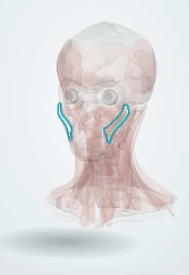
Muscular system · Head and neck
Zygomaticus major !
The zygomaticus major is a strap of muscle from the cheekbone to the corner of the mouth; it pulls the corner up and out to smile.
Official name: Zygomaticus major muscle
Opens in the Durgical app, no sign-up needed.
What it is
A strap from the cheekbone to the corner of the mouth.
What it does
Pulls the mouth corner up and out: the smile.
Key facts
- Origin
- Zygomatic bone (lateral)
- Insertion
- Angle of the mouth (modiolus)
- Action
- Pulls the mouth corner up and out (smile)
- Nerve
- Facial nerve (CN VII): zygomatic and buccal branches
- Blood supply
- Facial artery branches
Relations
- Superficial
- Malar fat pad and the SMAS of the face, then the skin of the cheek
- Deep
- Buccal fat pad, the buccinator, the masseter edge, and the facial artery and vein; the nerve branches enter from this side
- Medial
- Zygomaticus minor, running more medially into the upper lip
- Above
- Zygomatic bone, under the lower edge of the orbicularis oculi
- Below
- Modiolus at the angle of the mouth
How to find it
Asked to smile broadly, a band tightens from the zygomatic bone (the cheekbone) down to the mouth corner, lifting the corner up and out.
Examination
- Smiling broadly: the mouth corner does not rise on the weak side and the nasolabial fold flattens
- A genuine (Duchenne) smile adds the orbicularis oculi around the eye; in some central lesions an emotional smile is spared while a smile on request is weak
On imaging
Ultrasound and MRI show it as a band running from the zygomatic bone down to the corner of the mouth, superficial to the buccal fat pad and the facial vessels. It is mainly a landmark: ultrasound uses it to map the facial artery and vein before filler injections. In long-standing facial palsy it becomes thin. The muscle itself rarely needs imaging; facial nerve function is judged clinically.
Clinical notes
- Smile reanimation can join the masseteric nerve (V3) to a zygomatic branch, or transfer a free gracilis muscle to act in its place
- The zygomatic branches crossing the cheek are at risk in surgery over the zygoma and in deep-plane facelifts
- Botulinum toxin from neighbouring injections (masseter, lower lid) can spread to it and give an uneven smile
Anatomical variants
- A bifid zygomaticus major is a proposed cause of cheek dimples
Development
Second pharyngeal arch muscle; as this arch's muscle sheet splits into the separate muscles of expression, the facial nerve divides with it into the branches that reach each one.
Memory aid
Zygomaticus major makes the major smile; the minor only lifts the lip.
Test yourself
Think of the answer, then tap the question to see it.
What is the action of the zygomaticus major?
It pulls the mouth corner up and out: the main smiling muscle.
Which branches of the facial nerve supply the zygomaticus major?
The zygomatic and buccal branches, entering on its deep surface.
Why is the plane just superficial to the zygomaticus major safe in facelift dissection?
The facial artery and vein and the nerve branches lie deep to it.
Educational information, not medical advice. For any symptom, see a health professional.
Its names
- Official Latin (TA2)
- musculus zygomaticus major TA2 2079
- Official Spanish (DeCS)
- Músculo cigomático mayor
- English (TA2)
- Zygomaticus major muscle
- Also called
- musculus zygomaticus
Related
Sources: Terminologia Anatomica (TA2); Anatomy, Head and Neck: Facial Muscles; Anatomy, Head and Neck: Face; Anatomy of the Human Body (20th ed.): Development of the Muscles (Lea & Febiger (Gray's Anatomy, 1918), 1918) · adapted, Public domain.
Spotted a mistake?We are always improving for the community. Tell us what is wrong and our team will review it.Report a mistake
Representative model, for educational purposes only.