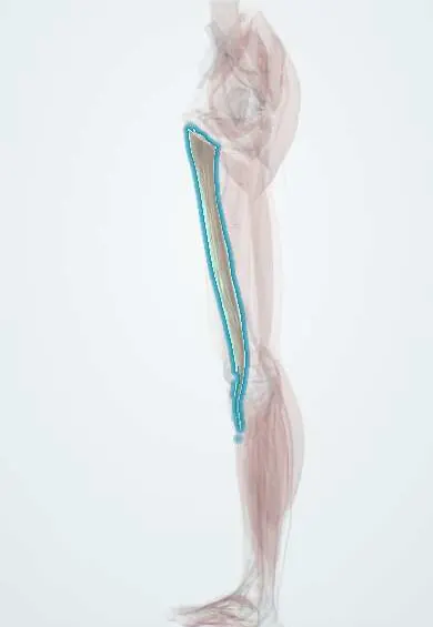
Gracilis !
The gracilis is a thin inner thigh muscle running from the pubic bone to the knee; it pulls the leg inward and helps bend the knee.
Official name: Gracilis muscle
Opens in the Durgical app, no sign-up needed.
What it is
A long, thin muscle on the inner thigh, from the pubic bone to the inner tibia just below the knee.
What it does
Pulls the leg inward and helps bend the knee.
Key facts
- Origin
- Body and inferior ramus of pubis
- Insertion
- Upper medial surface of tibia (pes anserinus)
- Action
- Adducts hip; flexes knee; medially rotates flexed knee
- Nerve
- Obturator nerve, anterior branch (L2–L3)
- Blood supply
- Medial circumflex femoral and deep femoral arteries; obturator artery
Relations
- Course
- From the pubis it runs straight down the medial side of the thigh as a thin strap, passes behind the medial femoral condyle and curves forward onto the upper medial tibia
- In front
- Adductor longus above; sartorius near the knee
- Deep
- Adductor brevis at its upper end, then adductor magnus
- Behind
- Semimembranosus and semitendinosus in the lower thigh
- Superficial
- Only fascia lata and the skin of the medial thigh, which makes it easy to reach and harvest
How to find it
With the hip abducted and the knee straight, resisting adduction makes its thin upper tendon stand out just behind the cord of the adductor longus near the pubis. At the knee, with the knee bent, it is the slimmer tendon felt just in front of the semitendinosus tendon on the inner back of the knee.
On imaging
On axial MRI it is the thin, most medial muscle of the thigh; at the knee its tendon lies between the sartorius and the semitendinosus in the pes anserinus. In groin pain, MRI can show a strain or avulsion of its origin on the pubis. Before ACL reconstruction, MRI or ultrasound can judge the size of the gracilis and semitendinosus tendons used for the graft.
Clinical notes
- Free functioning gracilis transfer, with its vessels from the deep femoral system and its motor branch from the anterior branch of the obturator nerve, can restore a smile in facial palsy or elbow flexion after brachial plexus injury
- Graciloplasty: the muscle is turned into the perineum to repair rectovaginal or rectourethral fistulas, or wrapped around the anal canal
- The pes anserine bursa lies between the three pes anserinus tendons and the medial collateral ligament; its inflammation causes pain a little below the medial joint line
Development
Formed from the ventral (flexor) muscle mass with the other adductors, hence its supply by the anterior branch of the obturator nerve.
Test yourself
Think of the answer, then tap the question to see it.
Which adductor is the only one to cross the knee?
The gracilis, the most superficial and most medial adductor; it runs from the pubis to the upper medial tibia (pes anserinus).
Which nerve supplies the gracilis?
The anterior branch of the obturator nerve (L2–L3); the same motor branch is used in free functioning gracilis transfer.
For which ligament reconstruction is the gracilis tendon harvested, and with which other tendon?
Anterior cruciate ligament (ACL) reconstruction, harvested with the semitendinosus tendon as a hamstring graft.
What can a free functioning gracilis muscle transfer restore?
A smile in facial palsy, or elbow flexion after brachial plexus injury; the muscle can be taken with little loss of function.
Educational information, not medical advice. For any symptom, see a health professional.
Its names
- Official Latin (TA2)
- musculus gracilis TA2 2635
- Official Spanish (DeCS)
- Músculo grácil DeCS D000071976
- English (TA2)
- Gracilis muscle
Related
Sources: Terminologia Anatomica (TA2); DeCS; Anatomy, Bony Pelvis and Lower Limb: Thigh Gracilis Muscle.
Spotted a mistake?We are always improving for the community. Tell us what is wrong and our team will review it.Report a mistake
Representative model, for educational purposes only.