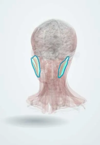
Muscular system · Head and neck
Masseter !
The masseter is the strongest chewing muscle, over the angle of the jaw; it closes the jaw and can ache from clenching or grinding the teeth.
Official name: Superficial part of masseter
Opens in the Durgical app, no sign-up needed.
What it is
The strongest chewing muscle; it is overworked in clenching and grinding (bruxism) and can ache with TMJ problems.
What it does
The strongest chewing muscle: closes the jaw.
Key facts
- Origin
- Zygomatic arch (superficial: anterior two thirds; deep: posterior part)
- Insertion
- Lateral ramus and angle of the mandible
- Action
- Elevates the mandible (closes the jaw); superficial part protrudes
- Nerve
- Masseteric nerve (V3)
- Blood supply
- Masseteric artery (maxillary artery), transverse facial artery
Relations
- Superficial
- Masseteric fascia, the parotid gland over its back part, the parotid duct, the transverse facial artery, the facial nerve branches, risorius, zygomaticus major and platysma
- Deep
- Ramus and coronoid process of the mandible, the insertion of the temporalis, and the masseteric nerve and vessels entering through the mandibular notch
- In front
- Buccinator and buccal fat pad, and the facial artery and vein crossing the mandible
- Behind
- Parotid gland and the retromandibular vein behind the ramus
- Above
- Zygomatic arch and zygomatic bone
How to find it
With the fingers on the angle of the jaw and the cheek in front of the ear, the person clenches the teeth: the muscle hardens and bulges under the fingers on both sides.
Examination
- Clenching while both sides are palpated: one soft side points to a lesion of the mandibular nerve on that side
- Jaw jerk: a tap on a finger on the chin with the mouth slightly open; a brisk reflex suggests bilateral upper motor neuron disease, such as pseudobulbar palsy
- Mouth opening measured between the incisors; reduced opening (trismus) suggests muscle spasm, infection of the masticator space or a joint problem
On imaging
Ultrasound and MRI show it as the thick muscle on the outer side of the mandibular ramus, with the parotid duct crossing its surface. Ultrasound compares both sides in benign masseteric hypertrophy and shows a masticator space abscess from a lower molar. Contrast CT or MRI map that abscess or a tumour of the masticator space, and CT shows fractures of the ramus and angle.
Common problems
- Clicking or popping when the disc slips forward and back.
- Locking (the jaw cannot open fully).
- Jaw pain linked to clenching or grinding (bruxism).
How it is diagnosed
Exam of jaw opening and the joint; MRI for the disc in persistent cases.
Treatment
- Soft diet, heat, jaw exercises and pain relief.
- A night guard (splint) for grinding.
- Joint washing (arthrocentesis) or surgery only rarely.
Clinical notes
- Benign masseteric hypertrophy, often with bruxism, gives a square lower face and must be told apart from a parotid tumour; botulinum toxin injection is one procedure used to reduce it
- Jaw pain on chewing that stops with rest (jaw claudication) is a classic symptom of giant cell arteritis
- Mandibular angle fractures are pulled apart or held together by the masseter and medial pterygoid sling, depending on the direction of the fracture line
- The masseteric nerve is a common donor for smile reanimation after facial palsy
Development
First pharyngeal arch muscle, so it is supplied by the mandibular nerve (V3), like all the chewing muscles.
Memory aid
Muscles of mastication come from the first arch and the mandibular nerve: «Mastication = Mandibular».
Related conditions
Test yourself
Think of the answer, then tap the question to see it.
What is the action and nerve supply of the masseter?
It elevates the mandible (closes the jaw); the superficial part protrudes it. Masseteric nerve (V3).
What is trismus, and what classic causes involve the masseter?
Limited mouth opening from spasm of the masseter and medial pterygoid: dental infection, pericoronitis of a lower wisdom tooth, or tetanus (lockjaw).
What does a brisk jaw jerk indicate?
An upper motor neuron lesion above the pons, such as pseudobulbar palsy; both arms of the reflex run in the mandibular nerve (V3).
Which duct crosses the outer surface of the masseter?
The parotid duct, which turns around the front edge of the masseter.
Jaw pain on chewing that stops with rest is a classic symptom of what?
Giant cell arteritis (jaw claudication).
Educational information, not medical advice. For any symptom, see a health professional.
Its names
- Official Latin (TA2)
- pars superficialis masseteris TA2 2106
- Official Spanish (DeCS)
- Músculo Masetero DeCS D008406
- English (TA2)
- Superficial part of masseter
Related
Sources: Terminologia Anatomica (TA2); DeCS; Anatomy, Head and Neck, Masseter Muscle; Anatomy, Head and Neck, Mastication Muscles.
Spotted a mistake?We are always improving for the community. Tell us what is wrong and our team will review it.Report a mistake
Representative model, for educational purposes only.