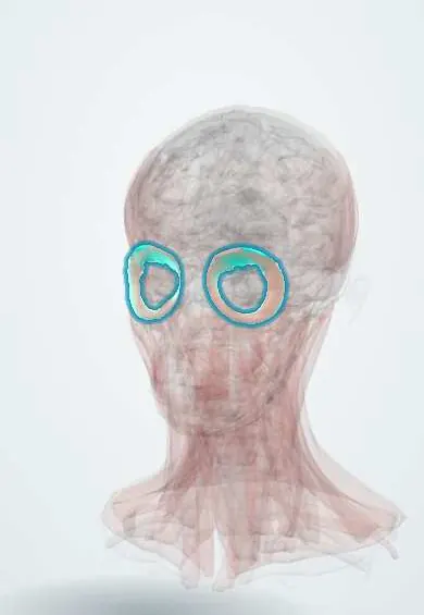
Muscular system · Head and neck
Orbicularis oculi !
The orbicularis oculi is the ring of muscle around the eye and inside the eyelids; it closes the eye to blink or squeeze, and fails to close it in Bell's palsy.
Official name: Orbital part of orbicularis oculi
Opens in the Durgical app, no sign-up needed.
What it is
A ring of muscle around the eye and inside the eyelids.
What it does
Closes the eye gently (blinking) or tightly (squeezing); it cannot close the eye in Bell's palsy.
Key facts
- Origin
- Medial orbital margin, medial palpebral ligament, lacrimal bone
- Insertion
- Skin around the orbit, lateral palpebral raphe
- Action
- Closes the eyelids (palpebral: blink; orbital: squeeze)
- Nerve
- Facial nerve (CN VII): temporal and zygomatic branches
- Blood supply
- Facial artery branches
Relations
- Superficial
- Thin eyelid skin, with almost no fat between them; thicker skin and fat over the orbital part
- Deep
- Orbital septum and tarsal plates of the eyelids; in the upper lid the levator palpebrae superioris; at the orbital rim the corrugator supercilii and suborbicularis fat
- Medial
- Medial palpebral ligament and the lacrimal sac of the tear drainage system, which the lacrimal part wraps behind
- Lateral
- Lateral palpebral raphe and the zygomatic bone
- Above
- Frontalis and the brow
- Below
- Origins of the levator labii superioris and zygomaticus minor and major
How to find it
Asked to close the eyes tightly, the ring contracts around the orbit, wrinkling the skin into crow's feet at the outer corner; a gentle blink moves only the lid part.
Examination
- Closing the eyes tightly against the examiner's fingers: the lid opens easily on a weak side, and the eyelashes stay visible (lashes not buried)
- Corneal reflex: touching the cornea gives no blink on the side of a facial nerve lesion, while the other eye still blinks (consensual), which separates it from a V1 lesion
- In a central lesion the eyes still close almost fully; in Bell palsy the eye cannot close
On imaging
Thin, so it is judged clinically rather than on images. On CT or MRI of the orbit it lies in the eyelids just under the skin, in front of the orbital septum. That septum is the key boundary: CT separates preseptal cellulitis, swelling only in front of it, from orbital cellulitis behind it, which needs urgent treatment and may hide an abscess. Ultrasound helps with eyelid lumps.
Clinical notes
- Exposure keratitis is the main risk in facial palsy; lid taping, lubrication, an upper-lid weight or tarsorrhaphy protect the cornea
- Paralysis of the lower lid lets it sag outward (paralytic ectropion) and tears run down the cheek, because the lacrimal pump fails
- Hemifacial spasm usually starts as twitching around the eye; it is often caused by a vessel pressing on the facial nerve near the brainstem
- Benign essential blepharospasm, a focal dystonia of this muscle, is treated with botulinum toxin injection
Development
A second pharyngeal arch muscle: the facial nerve enters this arch's muscle mass early and is pulled along as the mass spreads around the eye, which is why the eyelid closer depends on VII and not on III.
Related conditions
Test yourself
Think of the answer, then tap the question to see it.
What is the role of the orbicularis oculi in the corneal reflex?
It is the motor limb: touch is carried by V1 and the blink is made by the facial nerve through the orbicularis oculi.
What are the three parts of the orbicularis oculi and their actions?
Orbital (tight squeeze), palpebral (gentle blink) and lacrimal (pumps tears into the lacrimal sac).
What happens to the eye when the orbicularis oculi is paralysed?
The eye cannot close (lagophthalmos) and the cornea dries; on trying to close, the eyeball rolls up (Bell phenomenon).
Why do tears run down the cheek in lower-lid paralysis of the orbicularis oculi?
The lower lid sags outward (paralytic ectropion) and the lacrimal pump fails.
Educational information, not medical advice. For any symptom, see a health professional.
Its names
- Official Latin (TA2)
- pars orbitalis musculi orbicularis oculi TA2 2070
- Official Spanish (DeCS)
- Músculo orbicular del ojo DeCS D005152
- English (TA2)
- Orbital part of orbicularis oculi
Related
Sources: Terminologia Anatomica (TA2); DeCS; Anatomy, Head and Neck: Orbicularis Oculi Muscle; Anatomy of the Human Body (20th ed.): Development of the Muscles (Lea & Febiger (Gray's Anatomy, 1918), 1918) · adapted, Public domain.
Spotted a mistake?We are always improving for the community. Tell us what is wrong and our team will review it.Report a mistake
Representative model, for educational purposes only.