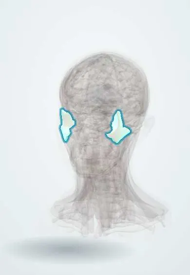
Skeletal system · Head and neck
Zygomatic bones !
The zygomatic bones are the cheekbones; they form the outer rim of the eye sockets, shape the cheeks and anchor the masseter chewing muscle.
Official name: Zygomatic bone
Opens in the Durgical app, no sign-up needed.
What it is
The cheekbones, forming the outer rim of the eye sockets.
What it does
Shape the cheek and anchor the masseter muscle.
Key facts
- Joints with
- Frontal, sphenoid, temporal (zygomatic arch), maxilla
- Landmarks
- Frontal, temporal and maxillary processes; lateral orbital rim; zygomatico-orbital, zygomaticofacial and zygomaticotemporal foramina (zygomatic nerve, V2)
Relations
- Medial
- Maxilla at the zygomaticomaxillary suture; the infraorbital foramen lies just below the orbital rim near this suture
- Above
- Zygomatic process of the frontal bone at the frontozygomatic suture
- Behind
- Zygomatic process of the temporal bone, forming the zygomatic arch; the greater wing of the sphenoid bone inside the orbit
- Deep
- Temporalis and the coronoid process of the mandible under the arch; the temporal and infratemporal fossae
- Superficial
- Zygomaticus major and minor arise from it; the masseter arises from the lower border of the arch
How to find it
The cheek prominence, the lateral orbital rim and the arch are all felt. Viewing both cheeks from above the head shows a flattened side, and a finger run along the infraorbital rim can find a step.
Examination
- Mouth opening: limited opening after a cheek injury suggests an arch fragment pressing on the coronoid process or temporalis
- Eye movements and double vision: restricted upward gaze suggests the orbital floor is also broken, with tissue trapped
- Sensation of the cheek, side of the nose and upper lip: reduced in infraorbital nerve injury
On imaging
Facial CT with coronal and 3D reconstructions is the standard; a submentovertex («jug-handle») radiograph shows the arch, and occipitomental views show the orbital rims and the maxillary sinuses.
Clinical notes
- Displaced complex fractures are reduced and fixed, often at the frontozygomatic suture, the infraorbital rim and the zygomaticomaxillary buttress
- An isolated depressed arch can be lifted through a small incision in the temporal hairline (Gillies approach)
- A subconjunctival haemorrhage whose back edge cannot be seen points to an orbital wall fracture
Development
Forms in membrane in the maxillary prominence of the first pharyngeal arch, usually from a single centre (some texts: up to three). In Treacher Collins syndrome it is small or missing, giving flat cheeks and down-slanting eyelids.
Related conditions
Test yourself
Think of the answer, then tap the question to see it.
What are the signs of a zygomaticomaxillary complex (tripod) fracture of the zygomatic bone?
Flat cheek, a step in the infraorbital rim, numb cheek and upper lip (infraorbital nerve), lateral subconjunctival haemorrhage and trismus.
Why can an isolated zygomatic arch fracture limit mouth opening?
The arch often caves in a V shape onto temporalis and the coronoid process of the mandible.
Through which approach is an isolated depressed zygomatic arch usually lifted?
The Gillies approach: a small incision in the temporal hairline.
Which tubercle on the zygomatic bone anchors the lateral canthal tendon?
The lateral orbital (Whitnall) tubercle, on the inside of the lateral orbital rim.
Educational information, not medical advice. For any symptom, see a health professional.
Its names
- Official Latin (TA2)
- os zygomaticum TA2 818
- Official Spanish (DeCS)
- Cigoma DeCS D015050
- English (TA2)
- Zygomatic bone
- Also called
- cheekbone · malar bone · zygoma
Sources: Terminologia Anatomica (TA2); DeCS; Anatomy, Head and Neck, Zygomatic; Anatomy, Head and Neck, Orbit Bones.
Spotted a mistake?We are always improving for the community. Tell us what is wrong and our team will review it.Report a mistake
Representative model, for educational purposes only.