Conditions · Obstetrics and gynecology
Asherman syndrome (intrauterine adhesions) !
Scar tissue forms inside the uterus, usually after a curettage. Periods become scant or stop, and getting or staying pregnant becomes harder.
Updated 2026-11-01
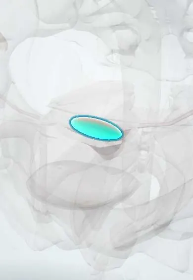
Structures involved
Tap one to see what it is and open it in 3D.
Why it happens
- Tissue repair: regeneration or scar; labile, stable and permanent cells
- Fibrosis in organs: TGF-β and the myofibroblast
- Uterus and the endometrial cycle
What it is
Acquired scarring within the uterine cavity, in which fibrous bands (synechiae) join the opposing walls after injury to the basal layer of the endometrium. Asherman syndrome is the name used when the adhesions cause symptoms such as menstrual disturbance, infertility or recurrent pregnancy loss.
Who gets it
True frequency is uncertain because many cases are mild. It is found more often in women who have had curettage after miscarriage or postpartum haemorrhage, and after repeated procedures. In Colombia and Latin America, where uterine evacuation for incomplete abortion and genital tuberculosis remain relevant, it is an important cause of secondary amenorrhoea and infertility.
Causes
- Uterine instrumentation, especially curettage of a pregnant or recently pregnant uterus (after miscarriage, retained products or postpartum haemorrhage) — the main cause
- Hysteroscopic surgery (removal of fibroids, septum or polyps)
- Caesarean section and uterine compression sutures
- Endometrial infection, including genital tuberculosis (a classic cause in endemic regions)
- Endometrial ablation and uterine artery embolisation
Risk factors
- Repeated curettage
- Curettage between 2 and 4 weeks after delivery or after missed miscarriage
- Retained products of conception
- Endometritis after delivery or abortion
- Multiple hysteroscopic procedures
- Genital tuberculosis
Pathophysiology
The endometrium regenerates each cycle from its basal layer. When this layer is damaged, especially in the low-oestrogen state after pregnancy, the raw surfaces of the anterior and posterior uterine walls heal into each other with fibrous tissue instead of new functional endometrium. The scarred, poorly vascularised cavity produces little or no menstrual flow, may block the cervix or tubal ostia, and offers a poor surface for implantation and placentation.
Classification
- Graded by extent of cavity involvement, type of adhesions (filmy or dense) and menstrual pattern (for example American Fertility Society and European Society for Gynaecological Endoscopy classifications)
Symptoms and signs
- Hypomenorrhoea (scant periods) or secondary amenorrhoea
- Cyclic pelvic pain without bleeding (blood trapped behind adhesions)
- Infertility
- Recurrent miscarriage
- Sometimes no symptoms
Diagnosis
- Clinical
- Suspected in a woman with scant or absent periods, infertility or recurrent loss after uterine instrumentation. Hormonal causes of amenorrhoea are excluded first (pregnancy test, FSH, prolactin, thyroid function); a negative progestin-plus-oestrogen withdrawal test points to an outflow or endometrial problem.
- Tests
- Pregnancy test and hormonal profile, which are normal. Endometrial biopsy and culture or PCR for tuberculosis when suspected.
- Imaging
- Hysteroscopy is the gold standard for diagnosis and treatment. Hysterosalpingography shows filling defects; saline sonohysterography, 3D transvaginal ultrasound or MRI can also help.
Differential diagnosis
- Pregnancy
- Hypothalamic amenorrhoea
- Hyperprolactinaemia
- Primary ovarian insufficiency
- Polycystic ovary syndrome
- Cervical stenosis
- Genital tuberculosis as underlying cause
Goal of treatment
Restore a normal uterine cavity and functional endometrium, prevent the adhesions from re-forming, and improve menstrual and reproductive outcomes.
Treatment
- Surgical
- Hysteroscopic adhesiolysis under direct vision, cutting the adhesions with minimal damage to healthy endometrium; severe cases may need several procedures.
- Procedure
- Measures to stop re-adhesion after surgery: a temporary intrauterine balloon or device, or adhesion barrier gels, followed by second-look hysteroscopy.
- Medical
- Oestrogen therapy after surgery (usually followed by a progestin) to promote endometrial regrowth; antituberculous treatment if genital tuberculosis is found.
- Follow-up
- Follow-up of menstrual pattern and reassessment of the cavity; future pregnancies are monitored for placental problems.
Complications
- Persistent infertility
- Recurrent miscarriage
- Placenta accreta spectrum and abnormal placentation in later pregnancies
- Preterm birth and fetal growth restriction
- Haematometra
- Uterine perforation during adhesiolysis
Prognosis
Mild and moderate adhesions respond well to hysteroscopic treatment, with restored menses and good chances of pregnancy. Dense, extensive adhesions or tuberculosis carry a high recurrence rate and poorer fertility outcomes.
Prevention
- Avoid unnecessary and repeated curettage
- Prefer medical management or vacuum aspiration for miscarriage where appropriate
- Use gentle hysteroscopic technique
- Treat endometritis promptly
Test yourself
Think of the answer, then tap the question to see it.
What is the most common cause of Asherman syndrome?
Curettage of a recently pregnant uterus (after miscarriage, retained products or postpartum haemorrhage).
Which endometrial layer must be damaged for adhesions to form?
The basal layer (stratum basalis), from which the endometrium regenerates.
What is the gold standard for diagnosis and treatment?
Hysteroscopy, with hysteroscopic adhesiolysis.
How do hormonal tests usually look in a woman with amenorrhoea from Asherman syndrome?
Normal (normal FSH, prolactin and thyroid tests); there is no withdrawal bleed after oestrogen plus progestin.
Which infection is a classic cause in endemic regions?
Genital (endometrial) tuberculosis.
Related conditions
- Female infertility
- Miscarriage (early pregnancy loss)
- Functional hypothalamic amenorrhoea
- Placenta accreta spectrum
- Tuberculosis
- Pelvic inflammatory disease
Educational information, not medical advice. For any symptom, see a health professional.
Sources: Focus on the Primary Prevention of Intrauterine Adhesions: Current Concept and Vision (International Journal of Molecular Sciences (PMC), 2021).
Spotted a mistake?We are always improving for the community. Tell us what is wrong and our team will review it.Report a mistake
Representative model, for educational purposes only.