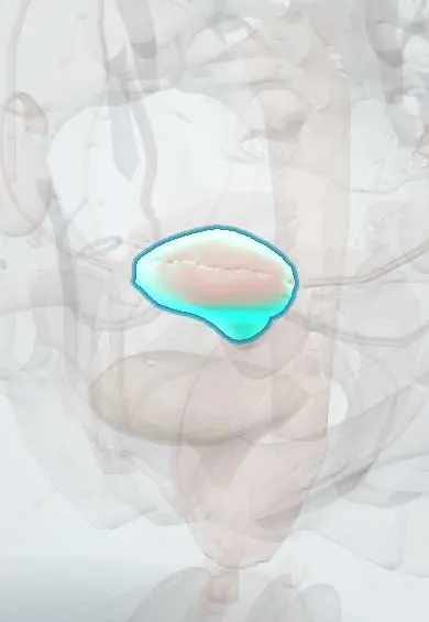
Reproductive system · Pelvis · Female
Body of uterus !
The body of the uterus is its main, muscular part, above the cervix; it is where a pregnancy implants and grows.
Opens in the Durgical app, no sign-up needed.
What it is
The main part of the uterus, with thick muscular front and back walls around a flat cavity.
What it does
Where a pregnancy implants and grows.
Key facts
- Where
- Between fundus and isthmus; front surface on the bladder (vesicouterine pouch), back surface toward the rectum (rectouterine pouch)
- Blood supply
- Uterine artery (internal iliac), anastomosing with the ovarian artery
- Nerve
- Uterovaginal plexus (from inferior hypogastric plexus): sympathetic T10–L1, parasympathetic S2–S4
- Function
- Implantation and growth of the pregnancy; contractions of labour
Relations
- In front
- Vesicouterine pouch and the upper surface of the urinary bladder
- Behind
- Rectouterine pouch, with loops of ileum or the sigmoid colon, and the rectum behind
- Lateral
- Broad ligament, with the uterine artery and uterine veins running up along its side
- Above
- Fundus of uterus, with the uterine tubes, round ligament of uterus and ovarian ligament attached at each upper corner
- Below
- Lower uterine segment and cervix
How to find it
On bimanual examination the body is held between the vaginal fingers and the abdominal hand to judge size, shape, mobility and tenderness; an anteverted uterus is felt mainly by the abdominal hand, a retroverted one through the posterior fornix. In pregnancy its size is compared with the dates.
On imaging
Transvaginal ultrasound in the sagittal plane shows position, size and the endometrial stripe. T2-weighted MRI separates the bright endometrium, the dark junctional zone and the intermediate outer myometrium, and is used to classify congenital anomalies.
Common problems
- Fibroids (benign muscle growths).
- Heavy or painful periods; adenomyosis.
- Endometrial cancer, usually after menopause.
- Prolapse (the uterus drops into the vagina).
How it is diagnosed
Pelvic examination and ultrasound (often transvaginal); hysteroscopy or a sample of the lining (biopsy) when needed; MRI for some cases.
Treatment
- Medicines or a hormonal intrauterine device for heavy bleeding.
- Removal of fibroids (myomectomy) or blocking their blood supply (embolisation).
- Hysterectomy (removal of the uterus) when other treatments are not enough or for cancer.
- Caesarean section to deliver a baby through the abdomen.
Clinical notes
- A uterus larger than expected for the dates suggests multiple pregnancy, fibroids, molar pregnancy or wrong dates
- Perforation during instrumentation (curettage, insertion of an intrauterine device) is more likely with a sharply flexed or retroverted uterus and soon after birth
- Total hysterectomy removes the body and the cervix; subtotal (supracervical) hysterectomy leaves the cervix
Anatomical variants
- Retroverted and retroflexed uterus: a normal variant in a minority of women (about one in five in many texts)
Development
The body forms from the fused paramesonephric (Müllerian) ducts; incomplete fusion gives a bicornuate uterus, and failure to resorb the midline wall a septate uterus.
Related conditions
- Postpartum haemorrhage
- Placental abruption
- Miscarriage (early pregnancy loss)
- Uterine fibroids (leiomyomas)
- Endometrial cancer
- Adenomyosis
- Abnormal uterine bleeding
- Dysmenorrhoea
- Pelvic organ prolapse
- Molar pregnancy (hydatidiform mole)
- Eclampsia
- Puerperal infection (postpartum endometritis and maternal sepsis)
- Rh alloimmunisation in pregnancy
- Chorioamnionitis (intra-amniotic infection)
- Septic and unsafe abortion
- Hyperemesis gravidarum
- Endometrial hyperplasia
- Gestational trophoblastic neoplasia (choriocarcinoma)
- Multiple pregnancy and twin–twin transfusion syndrome
- Breech and other malpresentations
- Stillbirth (intrauterine fetal death)
- Asherman syndrome (intrauterine adhesions)
- Müllerian anomalies (MRKH, septate uterus, imperforate hymen)
Test yourself
Think of the answer, then tap the question to see it.
What is the usual position of the body of the uterus?
Anteverted (cervix tipped forward on the vagina) and anteflexed (body bent forward on the cervix), resting on the bladder; retroversion is a common normal variant.
Why is the rectouterine pouch deeper than the vesicouterine pouch?
Behind the body of the uterus the peritoneum runs further down, over the posterior vaginal fornix; in front it turns onto the bladder at the isthmus.
What does incomplete fusion of the paramesonephric ducts produce, compared with failure to resorb the midline wall?
Incomplete fusion gives a bicornuate uterus; failure to resorb the midline wall gives a septate uterus.
What is the difference between total and subtotal hysterectomy?
Total hysterectomy removes the body of the uterus and the cervix; subtotal (supracervical) leaves the cervix.
Educational information, not medical advice. For any symptom, see a health professional.
Its names
- Official Latin (TA2)
- corpus uteri TA2 3502
- Official Spanish (DeCS)
- Cuerpo del útero DeCS D014599
- English (TA2)
- Body of uterus
Related
- Fundus of uterus
- Lower uterine segment
- Endometrium
- Myometrium
- Broad ligament
- Vesicouterine pouch
- Uterine artery
Sources: Terminologia Anatomica (TA2); DeCS; Anatomy, Abdomen and Pelvis: Uterus.
Spotted a mistake?We are always improving for the community. Tell us what is wrong and our team will review it.Report a mistake
Representative model, for educational purposes only.