Reproductive system · Pelvis · Female
Fundus of uterus !
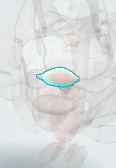
The fundus is the rounded top of the uterus, above where the tubes join; in pregnancy its height is measured to follow the baby's growth.
Opens in the Durgical app, no sign-up needed.
What it is
The rounded top of the uterus, above where the tubes join.
What it does
Its height above the pubic bone is measured in pregnancy to follow the baby's growth.
Key facts
- Where
- Rounded top of the uterus above the openings of the uterine tubes; normally anteverted and anteflexed over the bladder
- Blood supply
- Uterine artery (internal iliac), anastomosing with the ovarian artery
- Nerve
- Uterovaginal plexus (from inferior hypogastric plexus): sympathetic T10–L1, parasympathetic S2–S4
- Function
- Fundal height tracks pregnancy (at the umbilicus at about 20 weeks)
Relations
- Above
- Loops of ileum and the sigmoid colon; the greater omentum may reach it
- In front
- Upper surface of the urinary bladder when the uterus is anteflexed
- Lateral
- The cornua, with the uterine tubes, round ligament of uterus and ovarian ligament
- Below
- Body of uterus
How to find it
In pregnancy the fundus is found by walking the side of the hand down from the xiphoid until its upper edge is met; fundal height is then measured with a tape to the top of the pubic symphysis. After birth it is felt as a firm ball near the umbilicus that sinks a little each day.
Examination
- First Leopold manoeuvre: both hands palpate the fundus to tell which fetal pole lies there, a round hard head or a broad, softer breech
On imaging
On ultrasound, best in a coronal (3D) view, the outer contour of the fundus separates anomalies: a deep external cleft points to a bicornuate uterus, a smooth or flat contour with an inner septum to a septate uterus.
Common problems
- Fibroids (benign muscle growths).
- Heavy or painful periods; adenomyosis.
- Endometrial cancer, usually after menopause.
- Prolapse (the uterus drops into the vagina).
How it is diagnosed
Pelvic examination and ultrasound (often transvaginal); hysteroscopy or a sample of the lining (biopsy) when needed; MRI for some cases.
Treatment
- Medicines or a hormonal intrauterine device for heavy bleeding.
- Removal of fibroids (myomectomy) or blocking their blood supply (embolisation).
- Hysterectomy (removal of the uterus) when other treatments are not enough or for cancer.
- Caesarean section to deliver a baby through the abdomen.
Clinical notes
- A fundal height smaller or larger than expected leads to ultrasound for growth restriction, multiple pregnancy, abnormal amniotic fluid or wrong dates
- Uterine inversion: after delivery the fundus turns inside out through the cervix, classically with cord traction on a fundal placenta; the fundus can no longer be felt in the abdomen
- A high, soft fundus after birth signals uterine atony (see myometrium)
Anatomical variants
- Arcuate uterus: a shallow dip of the fundus into the top of the cavity with a normal outer contour; usually regarded as a variant of normal
Development
The fundus forms where the upper ends of the two paramesonephric (Müllerian) ducts fuse and the wall between them is resorbed; failure of fusion leaves a notched fundus (bicornuate uterus), while failure of resorption leaves an inner septum under a normal outer contour.
Related conditions
Test yourself
Think of the answer, then tap the question to see it.
Where is the uterine fundus felt at about 12 weeks and at about 36 weeks of pregnancy?
Just above the pubic symphysis at about 12 weeks; near the xiphoid process at about 36 weeks.
From about 24 weeks, how does symphysis–fundal height relate to gestational age?
The height in centimetres roughly equals the weeks of gestation, within about 2 cm.
What does the first Leopold manoeuvre assess at the uterine fundus?
Which fetal pole lies at the fundus: a round, hard head or a broad, softer breech.
In uterine inversion, what happens to the uterine fundus?
It turns inside out through the cervix after delivery, classically with cord traction on a fundal placenta, and can no longer be felt in the abdomen.
Educational information, not medical advice. For any symptom, see a health professional.
Its names
- Official Latin (TA2)
- fundus uteri TA2 3501
- Official Spanish (DeCS)
- Fondo del útero DeCS D014599
- English (TA2)
- Fundus of uterus
Related
Sources: Terminologia Anatomica (TA2); DeCS; Anatomy, Abdomen and Pelvis: Uterus.
Spotted a mistake?We are always improving for the community. Tell us what is wrong and our team will review it.Report a mistake
Representative model, for educational purposes only.