Conditions · Metabolism (biochemistry)
Congenital disorder of glycosylation type Ia (PMM2-CDG) !
The most common congenital disorder of glycosylation: proteins lack sugar chains, causing low tone, a small cerebellum and problems in many organs.
Updated 2026-11-01
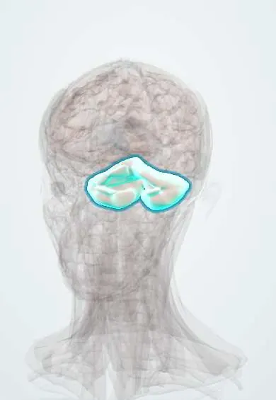
Structures involved
Tap one to see what it is and open it in 3D.
In biochemistry
What it is
An inherited defect of phosphomannomutase 2 (gene PMM2) that impairs N-linked glycosylation of proteins; it is the most frequent of the congenital disorders of glycosylation (CDG).
Who gets it
More than 800 affected people have been described worldwide; it is the most common CDG. Its frequency in Colombia is not known.
Causes
- Autosomal recessive variants in PMM2
Risk factors
- Carrier parents
- Affected sibling
Pathophysiology
PMM2 converts mannose 6-phosphate into mannose 1-phosphate, the precursor of GDP-mannose. Without enough GDP-mannose, the dolichol-linked oligosaccharide is not built properly and many proteins are under-glycosylated. Poorly glycosylated proteins fold badly, are degraded or work abnormally: clotting factors and anticoagulant proteins, hormones, transport proteins (transferrin) and brain proteins. The cerebellum is especially affected.
Classification
- Infantile multisystem form: hypotonia, failure to thrive, liver, heart and kidney involvement
- Late-infantile and childhood ataxia–intellectual disability form
- Adult stable disability form
Symptoms and signs
- Weak muscle tone and poor feeding in infancy
- Inverted nipples and abnormal fat pads on the buttocks
- Crossed eyes (strabismus) and abnormal eye movements
- Developmental delay, ataxia and later intellectual disability
- Stroke-like episodes and seizures
Diagnosis
- Clinical
- Infant with hypotonia, inverted nipples, abnormal fat distribution, strabismus and cerebellar signs.
- Tests
- Serum transferrin isoelectric focusing (type 1 pattern), liver enzymes, clotting factors (low factor XI, antithrombin, protein C and S), thyroid tests; PMM2 enzyme activity and gene testing confirm it.
- Imaging
- Brain MRI with cerebellar hypoplasia or atrophy; echocardiography for pericardial effusion or cardiomyopathy.
Treatment
- Rehabilitation
- Physiotherapy, occupational and speech therapy for motor and language delay
- Medical
- Treatment of seizures, thyroid dysfunction and clotting problems as they appear
- Follow-up
- Regular monitoring of liver, heart, kidneys, eyes and coagulation
- Conservative
- Nutritional support for feeding problems (thickened feeds or tube feeding when needed)
Complications
- Death in the first years in about a fifth of severely affected infants
- Thrombosis or bleeding
- Retinitis pigmentosa and progressive skeletal deformity
Prognosis
Severe infantile forms carry high early mortality; those who survive infancy often reach adulthood with stable disability.
Test yourself
Think of the answer, then tap the question to see it.
Which reaction does PMM2 catalyse?
Mannose 6-phosphate → mannose 1-phosphate, needed to make GDP-mannose for N-glycan synthesis.
Which screening test suggests a congenital disorder of glycosylation?
Isoelectric focusing of serum transferrin, which shows under-sialylated forms.
What are two typical physical findings of PMM2-CDG in infants?
Inverted nipples and abnormal fat pads, along with strabismus and hypotonia.
Why do patients have clotting abnormalities?
Clotting factors and natural anticoagulants (antithrombin, proteins C and S) are glycoproteins that are made poorly without proper glycosylation.
Related conditions
Educational information, not medical advice. For any symptom, see a health professional.
Sources: PMM2-congenital disorder of glycosylation: MedlinePlus Genetics (MedlinePlus, US National Library of Medicine, 2026); PMM2 gene: MedlinePlus Genetics (MedlinePlus, US National Library of Medicine, 2026); Textbook of Biochemistry for Medical Students, 6th ed. (Jaypee Brothers Medical Publishers (Vasudevan DM, Sreekumari S, Vaidyanathan K), 2011).
Spotted a mistake?We are always improving for the community. Tell us what is wrong and our team will review it.Report a mistake
Representative model, for educational purposes only.