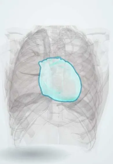
Pericardium !
The pericardium is the two-layered sac around the heart; it holds the heart in place and lets it beat without friction.
Opens in the Durgical app, no sign-up needed.
What it is
A tough double-layered sac around the heart, with a thin film of fluid between the layers.
What it does
Holds the heart in place, lets it beat without friction and limits sudden over-stretching.
Key facts
- Where
- Middle mediastinum, around heart and roots of great vessels; transverse and oblique sinuses
- Attaches
- Fibrous layer fused to central tendon of diaphragm (firmest in front) and great vessel adventitia; superior and inferior sternopericardial ligaments; weak vertebropericardial bands
- Function
- Fibrous layer limits overfilling; serous layers glide on fluid; phrenic nerves (C3–C5) carry its pain
Relations
- In front
- Body of the sternum and costal cartilages, tied by the sternopericardial ligaments; remains of the thymus above
- Behind
- Esophagus, Descending thoracic aorta and Azygos veins in the posterior mediastinum
- Lateral
- Mediastinal pleura and lungs, with the phrenic nerves and pericardiacophrenic vessels running between pleura and fibrous pericardium; these vessels, from the internal thoracic artery, are the main blood supply of the fibrous and parietal pericardium
- Below
- Central tendon of the Diaphragm, fused to the fibrous layer; the liver and stomach lie below
- Above
- Blends with the outer wall of the great vessels: Ascending aorta, Pulmonary trunk and superior vena cava; it rises highest on the aorta, near the origin of the brachiocephalic trunk, so the whole ascending aorta is inside the sac and its rupture causes tamponade
How to find it
Subxiphoid pericardiocentesis: the needle enters in the angle between the xiphoid process and the left costal margin, aimed toward the left shoulder, passing under the costal margin into the lowest part of the sac, now usually under echo guidance; this route avoids the pleura and the internal thoracic artery.
Examination
- Pericardial friction rub: a scratchy sound with up to three parts, heard best at the left sternal border with the person leaning forward; it points to pericarditis
- Pulsus paradoxus: a fall in systolic pressure of more than 10 mmHg on inspiration suggests tamponade
- Kussmaul sign: neck veins rise instead of fall on inspiration, typical of constrictive pericarditis
On imaging
Echocardiography is the first test for effusion and tamponade (diastolic collapse of the right atrium and ventricle, a full inferior vena cava). X-ray: a large effusion gives a globular, water-bottle heart. CT or MRI shows thickening and calcium in constriction; on CT, fluid in a recess (such as the superior aortic recess beside the ascending aorta) can mimic a node or a dissection.
Common problems
- Pericarditis: sharp chest pain, better leaning forward, often after a virus.
- Pericardial effusion: fluid builds up; if it squeezes the heart (tamponade) it is an emergency.
How it is diagnosed
ECG, echocardiogram, blood tests.
Treatment
- Anti-inflammatory medicines for pericarditis; draining the fluid with a needle (pericardiocentesis) for tamponade.
Clinical notes
- Pericarditis pain is sharp, worse lying flat and on breathing in, eased by sitting forward, and may spread to the trapezius ridge through the phrenic nerves; the ECG shows diffuse ST elevation with PR depression
- Constrictive pericarditis: a thick, sometimes calcified pericardium limits filling; tuberculosis remains an important cause in Colombia and other endemic settings
- Tamponade depends on speed: a small, fast bleed (trauma, after heart surgery, ventricular rupture) of only about 150–250 millilitres can compress the heart, while a slow effusion can stretch the sac to well over 1 L first; the normal sac holds about 15–50 millilitres of fluid
- Bare area: behind the lower left sternal edge and the 4th–5th left costal cartilages the sac touches the chest wall with no lung in front (cardiac notch); a parasternal needle or pericardial window here must avoid the internal thoracic vessels, about 1 cm from the sternum
Anatomical variants
- Congenital absence of the pericardium, usually partial and on the left, is rare; pericardial cysts, often at the right cardiophrenic angle, are benign
Development
The pericardial cavity comes from the intraembryonic coelom; the pleuropericardial folds that close it off carry the phrenic nerves, which explains their course on the pericardium.
Memory aid
Beck triad, the 3 Ds: Distant heart sounds, Distended neck veins, Decreased blood pressure.
Related conditions
- Systemic lupus erythematosus
- Mechanical complications of myocardial infarction
- Constrictive pericarditis
- Mediastinitis (acute and fibrosing)
- Blunt cardiac injury
- Acute pericarditis
- Cardiac tamponade
Test yourself
Think of the answer, then tap the question to see it.
Where is the transverse pericardial sinus?
Behind the aorta and pulmonary trunk and in front of the atria and superior vena cava; surgeons pass a finger or clamp through it.
What is Beck triad, and what does it indicate?
Low blood pressure, distended neck veins and muffled heart sounds: cardiac tamponade.
What is pulsus paradoxus?
A fall in systolic pressure of more than 10 mmHg on inspiration; it suggests tamponade.
How does pericarditis pain behave, and what does the ECG show?
Sharp, worse lying flat and breathing in, eased sitting forward, may spread to the trapezius ridge (phrenic nerves); diffuse ST elevation with PR depression.
What is the route of subxiphoid pericardiocentesis, and why is it used?
Between the xiphoid and the left costal margin, aimed at the left shoulder into the lowest part of the sac; it avoids the pleura and internal thoracic artery.
Educational information, not medical advice. For any symptom, see a health professional.
Its names
- Official Latin (TA2)
- pericardium; pericardium fibrosum; pericardium serosum TA2 3341
- Official Spanish (DeCS)
- Pericardio DeCS D010496
- English (TA2)
- Pericardium
Related
Sources: Terminologia Anatomica (TA2); DeCS; Anatomy, Thorax, Pericardium; Tratado de anatomía humana (9.ª ed.), tomo II, Libro IV Angiología, sección 1, cap. I Corazón (arts. VI–IX) y cap. II Pericardio.
Spotted a mistake?We are always improving for the community. Tell us what is wrong and our team will review it.Report a mistake
Representative model, for educational purposes only.