
Ocular rosacea
Rosacea of the eyes: gritty, red eyes and inflamed lids, sometimes before any skin signs. Severe cases scar the cornea.
Quick facts
- Key signs
- Burning · Red · Recurrent styes and chalazia
- Diagnosis
- Clinical
Red flags ↓
No doses: the clinician sets them by the current guideline.
What it is
Eye involvement in rosacea, a chronic inflammatory skin disease of the central face: meibomian gland dysfunction, blepharitis, dry eye and recurrent chalazia, and in severe cases keratitis with vessels, thinning or perforation. Eye signs come before skin signs in up to one in five patients, and children can be affected.
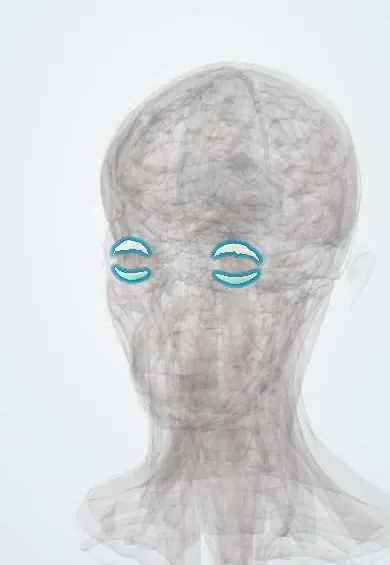
See it in 3D
Who gets it
More than half of people with skin rosacea have some eye involvement. It affects adults aged 30 to 60, men and women about equally for eye disease, and is often missed in darker skin. Childhood ocular rosacea presents with recurrent chalazia and keratitis.
Causes
- Dysregulated innate immunity (cathelicidin and other peptides)
- Neurovascular dysregulation with flushing
- Demodex mites and their bacteria
- Genetic predisposition
Risk factors
- Facial rosacea
- Fair skin
- Triggers: heat, sun, alcohol, spicy food, stress
Pathophysiology
Abnormal innate immune activity and increased matrix metalloproteinases in the tear film inflame the meibomian glands and lid margin. Thickened meibum destabilises the tear film, causing evaporative dry eye. Inflammatory mediators reach the cornea, causing peripheral vessels, infiltrates and, through collagenase activity, thinning.
Classification
- Mild: lid margin telangiectasia, meibomian dysfunction, mild irritation
- Moderate: chalazia, conjunctival redness, punctate keratopathy
- Severe: corneal vascularisation, infiltrates, ulceration, thinning or perforation; scleritis or episcleritis
Symptoms and signs
- Burning, gritty, watery or dry eyes
- Red, thickened lid margins with telangiectasia and capped meibomian glands
- Recurrent styes and chalazia
- Light sensitivity and blurred vision when the cornea is involved
- Facial flushing, erythema, papules and pustules (may be absent)
Diagnosis
- Clinical
- Clinical diagnosis from lid margin signs, meibomian dysfunction and facial rosacea; slit-lamp examination of the cornea with fluorescein.
- Tests
- Tear break-up time and other dry eye tests; culture if keratitis may be infectious; biopsy of a recurrent chalazion in older adults to exclude carcinoma.
- Imaging
- Meibography to show gland dropout where available.
Differential diagnosis
- Staphylococcal or seborrhoeic blepharitis
- Dry eye disease of other causes
- Sebaceous gland carcinoma (unilateral, recurrent chalazion)
- Herpes simplex keratitis
- Allergic conjunctivitis
Gritty eyes with inflamed lids: which one is it?
| Feature | Ocular rosacea | Staphylococcal blepharitis | Sebaceous gland carcinoma |
|---|---|---|---|
| Lid margin | Telangiectasia, capped meibomian glands, thick oil | Crusts and collarettes at the lash bases | Thickened lid, lash loss, a nodule that keeps returning |
| Laterality | Both eyes | Both eyes | One eye |
| Skin | Facial flushing, papules, pustules (may be absent) | Usually normal | Usually normal |
| Cornea | Vessels, infiltrates, thinning in severe cases | Marginal infiltrates possible | Usually spared; can mimic chronic one-sided conjunctivitis |
| Key step | Long-term lid care; oral tetracycline or macrolide | Lid hygiene | Biopsy |
Goal of treatment
Control lid and meibomian inflammation, protect the cornea and treat the skin disease together with dermatology.
Treatment
- Conservative
- Warm compresses, lid massage and lid hygiene; preservative-free lubricants; avoid known triggers.
- Medical
- Oral doxycycline or another tetracycline at anti-inflammatory levels in adults; oral macrolide (azithromycin or erythromycin) in children and pregnancy.
- Medical
- Topical azithromycin, metronidazole or ivermectin for the lids and skin; topical cyclosporine; short courses of topical corticosteroid for keratitis under ophthalmology follow-up.
- Procedure
- Intense pulsed light or thermal pulsation for meibomian disease; amniotic membrane, tissue glue or grafting for corneal thinning or perforation.
Complications
- Corneal scarring and vascularisation
- Corneal thinning and perforation
- Chronic dry eye
- Recurrent chalazia
Common traps
- No facial rash means no rosacea: eye signs come first in up to about one in five patients and skin signs are easily missed on darker skin, so lid telangiectasia with meibomian disease is enough to suspect it
- Doxycycline is given in ocular rosacea to kill an infection: it is used for its anti-inflammatory and anti-collagenase effect; macrolides replace it in children and pregnancy
- Recurrent chalazia in a child are just bad luck: they are a classic presentation of childhood ocular rosacea, which can also vascularise and scar the cornea
- A chalazion that keeps returning in the same spot in an older adult is rosacea: a recurrent one-sided chalazion is biopsied to exclude sebaceous gland carcinoma
- Ocular rosacea is only a nuisance: corneal vascularisation, thinning and perforation can follow, so a painful red eye with an infiltrate needs prompt ophthalmology review
Prognosis
A chronic, relapsing condition that is usually well controlled with ongoing lid care and intermittent oral therapy; severe corneal disease can permanently reduce vision.
Prevention
- Daily lid hygiene
- Sun protection and avoidance of personal triggers
Test yourself
Think of the answer, then tap the question to see it.
A woman with facial flushing has burning eyes, lid-margin telangiectasia and capped meibomian glands. Why are her eyes dry?
Inflamed meibomian glands make thick, poor-quality oil, so the tear film evaporates quickly: evaporative dry eye from ocular rosacea.
Why does doxycycline help ocular rosacea when it is not an infection?
It reduces inflammation and inhibits matrix metalloproteinases and collagenase, which calms the glands and protects the cornea from thinning.
A 7-year-old has recurrent chalazia and peripheral corneal vessels with infiltrates. What is the diagnosis, and which oral drug class?
Childhood ocular rosacea; a macrolide such as azithromycin or erythromycin, because tetracyclines stain developing teeth.
A 70-year-old has a chalazion on the same lid for the third time. What next?
Biopsy, to exclude sebaceous gland carcinoma, which can masquerade as a recurrent chalazion or chronic one-sided blepharitis.
Which finding in ocular rosacea calls for urgent ophthalmology review?
A painful red eye with a corneal infiltrate, thinning or reduced vision, because the cornea can ulcerate and perforate.
Why it happens
In dermatology
Connections
Educational information, not medical advice. For any symptom, see a health professional.
Sources: Ocular Rosacea (EyeWiki, American Academy of Ophthalmology, 2026); Rosacea (StatPearls Publishing (NCBI Bookshelf), 2023).
Spotted a mistake?We are always improving for the community. Tell us what is wrong and our team will review it.Report a mistake
Representative model, for educational purposes only.