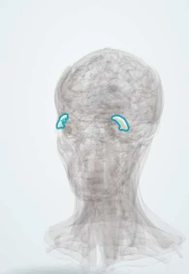
Lacrimal gland !
The lacrimal gland is an almond-sized gland in the upper outer corner of the eye socket; it makes the watery part of tears.
Opens in the Durgical app, no sign-up needed.
What it is
An almond-sized gland in the upper outer corner of the eye socket.
What it does
Makes the watery part of tears.
Key facts
- Where
- Upper outer orbit (lacrimal fossa of the frontal bone)
- Blood supply
- Lacrimal artery (ophthalmic)
- Nerve
- Parasympathetic: VII (greater petrosal nerve -> pterygopalatine ganglion -> zygomatic/lacrimal nerves)
- Function
- Secretes the watery tears
Relations
- Above
- Lacrimal fossa on the underside of the orbital part of the frontal bone, just behind the upper outer orbital rim
- Below
- Lateral rectus, along whose upper border the lacrimal artery and lacrimal nerve reach the gland
- Medial
- Levator palpebrae superioris, whose lateral horn passes between the two lobes
- In front
- Orbital septum and the lateral upper eyelid; the palpebral lobe lies under the conjunctiva of the upper lateral fornix
- Behind
- Orbital fat
How to find it
Normally it is not felt. Lifting the outer part of the upper lid while the person looks down and toward the nose shows the palpebral lobe as a pinkish, lobulated mass in the upper lateral fornix. An enlarged gland can be felt under the upper outer orbital rim and gives the upper lid an S-shaped curve.
Examination
- Schirmer test: a filter-paper strip hooked over the lower lid margin for 5 minutes; a short wet length (under about 5 mm; some texts use 10 mm) points to low aqueous tear production
- Tear break-up time with fluorescein and corneal staining: a fast break-up with normal Schirmer suggests evaporative (meibomian) dry eye rather than low tear output
On imaging
CT and MRI of the orbit show it in the upper outer orbit. Diffuse enlargement of both glands suggests inflammation, lymphoma or sarcoidosis; a well-defined mass that remodels the bone of the fossa fits a pleomorphic adenoma, while bone erosion with pain suggests a malignant tumour such as adenoid cystic carcinoma.
Clinical notes
- A facial nerve (VII) lesion at or above the geniculate ganglion, or of the greater petrosal nerve, reduces tearing on that side; lesions further down spare it, which helps locate the lesion
- "Crocodile tears": after a facial palsy, regrowing salivary fibres can reach the gland, so the eye waters while eating
- Dry eye from low tear output: Sjögren syndrome (with a dry mouth), ageing and gland damage; it causes gritty, burning eyes and, paradoxically, bouts of reflex watering
- Dacryoadenitis gives a tender, swollen upper outer lid with an S-shaped margin; a lacrimal gland tumour pushes the eye down and inward
Development
It grows as buds of the conjunctival epithelium in the upper lateral fornix and is not fully mature at birth, so newborns often cry with few tears during the first weeks.
Related conditions
Test yourself
Think of the answer, then tap the question to see it.
What divides the lacrimal gland into two lobes, and where do its ducts open?
The lateral horn of the levator palpebrae aponeurosis; the ducts open into the upper lateral conjunctival fornix.
Why can removing or biopsying the palpebral lobe of the lacrimal gland leave a dry eye?
The ducts of the orbital lobe pass through the palpebral lobe, so the whole gland can be cut off.
What is the parasympathetic pathway to the lacrimal gland?
Facial nerve (VII), greater petrosal nerve, pterygopalatine ganglion, then the zygomatic and lacrimal nerves.
What are «crocodile tears»?
After a facial palsy, regrowing salivary fibres reach the lacrimal gland, so the eye waters while eating.
What does the Schirmer test measure, and what result points to low tear production?
Aqueous tear output with a filter-paper strip over the lower lid for 5 minutes; a wet length under about 5 mm (some texts 10 mm).
Educational information, not medical advice. For any symptom, see a health professional.
Its names
- Official Latin (TA2)
- glandula lacrimalis TA2 6846
- Official Spanish (DeCS)
- Glándula lagrimal DeCS D007765
- English (TA2)
- Lacrimal gland
Related
Sources: Terminologia Anatomica (TA2); DeCS; Anatomy, Head and Neck: Eye Lacrimal Gland.
Spotted a mistake?We are always improving for the community. Tell us what is wrong and our team will review it.Report a mistake
Representative model, for educational purposes only.