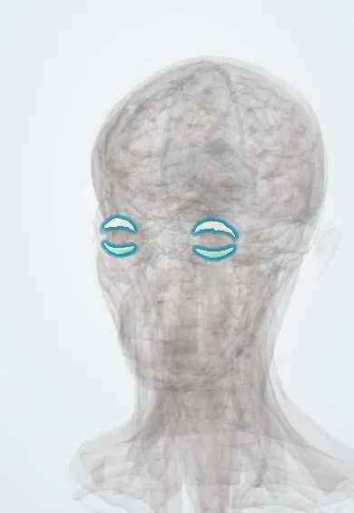
Eyelids !
The eyelids are folds of skin and muscle stiffened by plates of dense tissue; they protect the eye and spread the tears with each blink.
Official name: Superior tarsus
Opens in the Durgical app, no sign-up needed.
What it is
Folds of skin and muscle stiffened by plates of dense tissue (tarsi).
What it does
Protect the eye and spread the tears with each blink.
Key facts
- Where
- In front of the eye; tarsal plates give them shape
- Blood supply
- Palpebral arcades (ophthalmic and facial arteries)
- Nerve
- Opening: III (+ sympathetic Müller muscle); closing: VII; sensory V1/V2
- Function
- Protect the eye and spread tears
Relations
- Superficial
- Very thin skin over loose areolar tissue without fat, so blood and oedema collect easily (a black eye)
- Deep
- Palpebral conjunctiva lining the tarsus, which turns at the fornices onto the eyeball; the lid margins rest on the cornea
- Contents
- Tarsal plates with the tarsal (meibomian) glands; the palpebral part of orbicularis oculi; in the upper lid, the levator palpebrae aponeurosis and the superior tarsal muscle; lashes with the glands of Zeis and Moll
- Medial
- Medial canthus: the lacrimal puncta and canaliculi, and the medial palpebral ligament in front of the lacrimal sac
- Lateral
- Lateral palpebral ligament (lateral canthal tendon), anchored to the orbital tubercle of the zygomatic bone
How to find it
The upper lid crease lies a few millimetres above the margin, where levator fibres reach the skin; the tarsal plate is felt as a firm sheet inside each lid. On the margin, the lashes sit in front, the grey line in the middle and the row of meibomian gland openings behind it; the puncta are small openings on the medial end of each margin.
Examination
- Upper lid eversion over a cotton swab placed at the upper edge of the tarsus: shows a foreign body hidden under the lid or the papillae of allergic conjunctivitis
- Forced lid closure against the examiner's fingers tests orbicularis oculi and the facial nerve (VII); weak closure that leaves the cornea exposed (lagophthalmos) is typical of Bell palsy
- Lower lid distraction (snap-back) test: a lid that pulls far from the globe and returns slowly shows horizontal laxity, the basis of age-related ectropion and entropion
On imaging
Examined clinically; imaging is used for depth. Contrast CT of the orbits separates preseptal cellulitis, which stays in front of the orbital septum, from orbital cellulitis behind it, with fat stranding, a subperiosteal abscess or proptosis. It is chosen when there is pain on eye movement, reduced vision or proptosis. MRI shows how far an eyelid tumour has spread into the orbit.
Clinical notes
- Chalazion: a painless, firm lump from a blocked meibomian gland (a chronic granuloma); hordeolum (stye): an acute, painful infection of a lash gland (external) or a meibomian gland (internal)
- Blepharitis: chronic inflammation of the lid margin with crusting and meibomian gland dysfunction, a common cause of burning, gritty eyes
- Ectropion (margin turned out) causes watering and exposure; entropion (turned in) rubs the lashes on the cornea. Both are mostly age-related laxity; ectropion also follows facial palsy or skin scarring
- A laceration medial to the puncta may cut the lacrimal canaliculus (dog bites, blows); it is repaired over a stent so that tears keep draining
- The lower lid is a common site of basal cell carcinoma; a chalazion that keeps coming back in an older adult is biopsied to exclude sebaceous gland carcinoma
Anatomical variants
- In many East Asian eyelids the levator fibres reach the skin lower and the fat in front of the aponeurosis sits lower, so the lid crease is low or absent (single eyelid)
- Epicanthal folds: a vertical skin fold over the medial canthus, normal in infants and in many East Asian people and also seen in Down syndrome; covering the inner white of the eye, it can make the eyes look crossed (pseudostrabismus)
Development
The lid folds grow over the developing eye and fuse early in the fetal period; lashes and glands form while they are closed, and the lids separate again around the sixth to seventh month.
Related conditions
- Chalazion and hordeolum (stye)
- Basal cell carcinoma
- Stevens–Johnson syndrome and toxic epidermal necrolysis
- Infantile hemangioma
- Horner syndrome
- Chemical eye injury
- Corneal abrasion and ocular foreign body
- Herpes zoster ophthalmicus
- Thyroid eye disease (Graves orbitopathy)
- Trachoma
- Dacryocystitis and nasolacrimal duct obstruction
- Dry eye disease
Test yourself
Think of the answer, then tap the question to see it.
What are the layers of the eyelid from front to back?
Skin, orbicularis oculi, orbital septum, tarsal plate (with the levator aponeurosis and superior tarsal muscle in the upper lid), then palpebral conjunctiva.
What does the grey line on the eyelid margin mark?
The plane between the anterior lamella (skin, orbicularis oculi) and the posterior lamella (tarsus, conjunctiva), used to split the lid in surgery.
Which eyelid structure separates preseptal infections from orbital ones?
The orbital septum.
How does a chalazion differ from a hordeolum (stye)?
Chalazion: a painless, firm lump from a blocked meibomian gland (chronic granuloma); hordeolum: an acute, painful infection of a lash or meibomian gland.
Why is a recurring chalazion in an older adult biopsied?
To exclude sebaceous gland carcinoma.
Educational information, not medical advice. For any symptom, see a health professional.
Its names
- Official Latin (TA2)
- tarsus superior TA2 6827
- Official Spanish (DeCS)
- Párpados DeCS D005143
- English (TA2)
- Superior tarsus
- Also called
- lids
Related
Sources: Terminologia Anatomica (TA2); DeCS; Anatomy, Head and Neck: Eyelid.
Spotted a mistake?We are always improving for the community. Tell us what is wrong and our team will review it.Report a mistake
Representative model, for educational purposes only.